Main Model
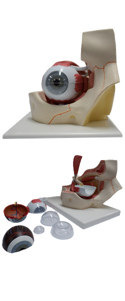
Uvea : 3 Iris with pupil

Vascular Layer of Eyeball
The middle vascular layer of the eyeball (also called the
uvea or uveal tract) consists of the choroid, ciliary body, and
iris. The choroid, a dark reddish brown layer
between the sclera and retina, forms the largest part of the
vascular layer of the eyeball and lines most of the sclera. Within this pigmented and dense vascular bed, larger
vessels are located externally (near the sclera). The finest vessels (the capillary lamina of the choroid, or choriocapillaris, an extensive capillary bed) are innermost, adjacent to
the avascular light-sensitive layer of the retina, which it supplies with oxygen and nutrients. Engorged with blood in life
(it has the highest perfusion rate per gram of tissue of all vascular beds of the body), this layer is responsible for the "red eye" reflection that occurs in flash photography. The choroid attaches firmly to the pigment layer of the retina, but can
easily be stripped from the sclera. The choroid is continuous anteriorly with the ciliary body.
The ciliary body, is a ring-like thickening of the layer posterior to the corneoscleral junction, which is muscular as well
as vascular. It connects the choroid
with the circumference of the iris. The ciliary body provides
attachment for the lens. The contraction and relaxation of the circularly arranged smooth muscle of the ciliary body controls
the thickness, and therefore the focus, of the lens. Folds on
the internal surface of the ciliary body, the ciliary processes,
secrete aqueous humor. Aqueous humor fills the anterior
segment of the eyeball, the interior of the eyeball anterior
to the lens, suspensory ligament, and ciliary body.
The iris, which literally lies on the anterior surface of
the lens, is a thin contractile diaphragm with a central aperture, the pupil, for transmitting light. When a person is awake, the size of the pupil varies
continually to regulate the amount of light entering the eye. Two involuntary muscles control the size of the
pupil: the parasympathetically stimulated, circularly arranged sphincter pupillae decreases its diameter (constrict or contracts the pupil, pupillary miosis), and the sympathetically
stimulated, radially arranged dilator pupillae increases
its diameter (dilates the pupil). The nature of the pupillary
responses is paradoxical: sympathetic responses usually occur
immediately, yet it may take up to 20 minutes for the pupil to dilate
in response to low lighting, as in a darkened theater. Parasympathetic responses are typically slower than sympathetic
responses, yet parasympathetically stimulated papillary
constriction is normally instantaneous. Abnormal sustained
pupillary dilation (mydriasis) may occur in certain diseases or as a result of trauma or the use of certain drugs.