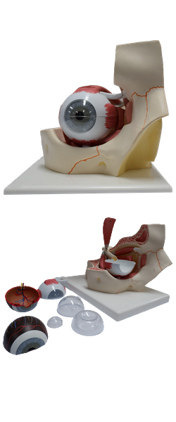
Main Model
Inner layer of eyeball : 9 Retina

Inner Layer of Eyeball
The inner layer of the eyeball is the retina. It is the sensory neural layer of the eyeball. Grossly, the retina consists of two functional parts with distinct locations: an optic part and a non-visual retina. The optic part
of the retina is sensitive to visual light rays and has two layers: a neural layer and pigmented layer. The neural layer
is light receptive. The pigmented layer consists of a single
layer of cells that reinforces the light-absorbing property of
the choroid in reducing the scattering of light in the eyeball.
The non-visual retina is an anterior continuation of the pigmented layer and a layer of supporting cells. The non-visual
retina extends over the ciliary body (ciliary part of retina)
and the posterior surface of the iris (iridial part of retina),
to the pupillary margin.
Clinically, the internal aspect of the posterior part of the
eyeball, where light entering the eyeball is focused, is referred
to as the fundus of the eyeball (ocular fundus). The retina
of the fundus includes a distinctive circular area called the
optic disc (optic papilla), where the sensory fibers and vessels conveyed by the optic nerve (CN II) enter the eyeball. Because it contains no photoreceptors, the optic disc is insensitive to light. Consequently,
this part of the retina is commonly called the blind spot.
Just lateral to the optic disc is the macula of the retina or
macula lutea (Latin yellow spot). The yellow color of the macula is
apparent only when the retina is examined with red-free light.
The macula is a small oval area of the retina with special photoreceptor cones that is specialized for acuity of vision. It is not
normally observed with an ophthalmoscope (a device for viewing the interior of the eyeball through the pupil). At the center
of the macula, a depression, the fovea centralis (Latin central pit), is the area of most acute vision. The fovea is approximately
1.5 mm in diameter; its center, the foveola, does not have the
capillary network visible elsewhere deep to the retina.
The optic part of the retina terminates anteriorly along
the ora serrata (Latin serrated edge), the irregular posterior
border of the ciliary body. Except
for the cones and rods of the neural layer, the retina is supplied by the central artery of the retina, a branch of the
ophthalmic artery. The cones and rods of the outer neural
layer receive nutrients from the capillary lamina of the choroid, or choriocapillaris. It has the finest vessels of the inner surface of the
choroid, against which the retina is pressed. A corresponding system of retinal veins unites to form the central vein
of the retina.