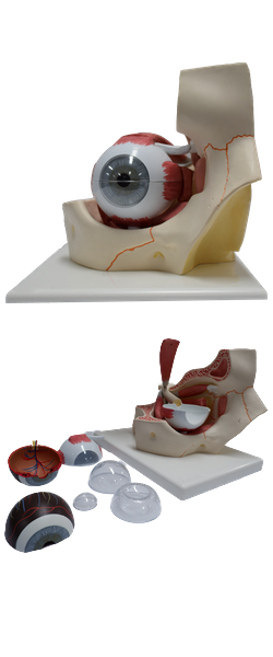
Main Model
Fibrous layer of eyeball : 2 Sclera

Fibrous Layer of Eyeball
The fibrous layer of the eyeball is the external fibrous skeleton of the eyeball, providing shape and resistance. The sclera is the tough opaque part of the fibrous layer (coat) of the eyeball, covering the posterior five sixths of the eyeball. It provides attachment for both the extrinsic (extra-ocular) and intrinsic muscles of the eye. The anterior part of the sclera is visible through the transparent bulbar conjunctiva as "the white of the eye". The cornea is the transparent part of the fibrous layer covering the anterior one sixth of the eyeball. The convexity of the cornea is greater than that of the sclera, and so it appears to protrude from the eyeball when viewed laterally.
The two parts of the fibrous layer differ primarily in terms of the regularity of arrangement of the collagen fibers of which they are composed and the degree of hydration of each. While the sclera is relatively avascular, the cornea is completely avascular, receiving its nourishment from capillary beds around its periphery and fluids on its external and internal surfaces (lacrimal fluid and aqueous humor, respectively). Lacrimal fluid also provides oxygen absorbed from the air.
The cornea is highly sensitive to touch, its innervation is provided by the ophthalmic nerve (CN V1). Even very small foreign bodies (e.g., dust particles) elicit blinking, flow of tears, and sometimes severe pain. Its nourishment is derived from the capillary beds at its periphery, the aqueous humor, and lacrimal fluid. The latter also provides oxygen absorbed from air. Drying of the corneal surface may cause ulceration.
The corneal limbus is the angle formed by the intersecting curvatures of sclera and cornea at the corneoscleral junction. The junction is a 1-mm-wide, gray, translucent circle that includes numerous capillary loops involved in nourishing the avascular cornea.