Main Model
Orbit : 19 Optic chiasm

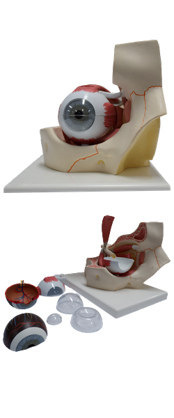
Optic Nerve (CN II)
Function: Special sensory (special somatic afferent) - that is, the special sense of vision.
Although officially nerves by convention, the optic nerves (CN II) develop in a completely different manner from the other cranial nerves. The structures involved in receiving and transmitting optical stimuli (the optical fibers and neural retina, together with the pigmented epithelium of the eyeball) develop as evaginations of the diencephalon. The optic nerves are paired, anterior extensions of the forebrain (diencephalon) and are therefore actually CNS fiber tracts formed by axons of retinal ganglion cells. In other words, they are third order neurons, with their cell bodies located in the retina.
The optic nerves are surrounded by extensions of the cranial meninges and subarachnoid space, which is filled with cerebrospinal fluid (CSF). The meninges extend all the way to the eyeball. The central artery and vein of the retina traverse the meningeal layers and course in the anterior part of the optic nerve. Cranial nerve II begins where the unmyelinated axons of retinal ganglion cells pierce the sclera (the opaque part of the external fibrous coat of the eyeball) and become myelinated, deep to the optic disc.
The nerve passes posteromedially in the orbit, exiting through the optic canal to enter the middle cranial fossa, where it forms the optic chiasm. Here, fibers from the nasal (medial) half of each retina decussate in the chiasm and join uncrossed fibers from the temporal (lateral) half of the retina to form the optic tract.
The partial crossing of optic nerve fibers in the chiasm is a requirement for binocular vision, allowing depth-of-field perception (three-dimensional vision). Thus fibers from the right halves of both retinas form the right optic tract. The decussation of nerve fibers in the chiasm results in the right optic tract conveying impulses from the left visual field and vice versa. The visual field is what is seen by a person who has both eyes wide open and who is looking straight ahead. Most fibers in the optic tracts terminate in the lateral geniculate bodies of the thalamus. From these nuclei, axons are relayed to the visual cortices of the occipital lobes of the brain.