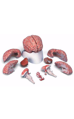
Main Model
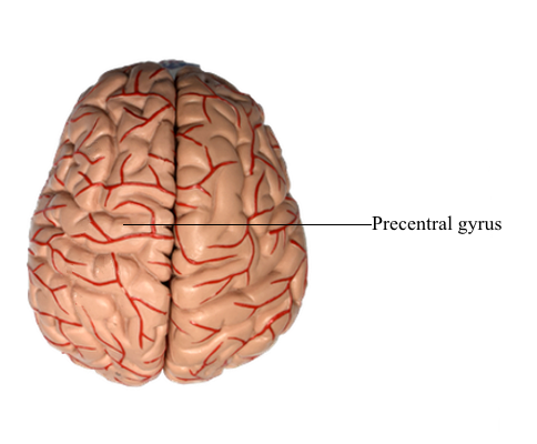
Telencephalon : 6 Precentral gyrus
Frontal Lobe
The lateral surface of the frontal lobe is divided by inferior and superior frontal sulci into inferior, middle, and superior frontal gyri, the last folding onto the medial aspect of the hemisphere. The inferior frontal gyrus is divided into a pars opercularis, pars triangularis, and pars orbitalis. The anterior (orbital) surface of the frontal lobe is composed of the gyrus rectus, the olfactory sulcus, and a series of orbital gyri. The most rostral point of this lobe is the frontal pole of the brain.
The olfactory bulb and tract, which relay sensory information, lie on the anterior surface of the frontal lobe in the olfactory sulcus. At the point where the olfactory tract attaches to the hemisphere, it bifurcates into medial and lateral striae. The triangle formed by this bifurcation is called the olfactory trigone. Immediately caudal to this trigone, the surface of the hemisphere is characterized by numerous small holes formed by vessels (lenticulostriate arteries) as they enter the brain; this is the anterior perforated substance. The olfactory tract and striae and the cell groups associated with the anterior perforated substance are functionally related to the limbic system.
The precentral gyrus is continuous on the medial surface of the hemisphere with the anterior paracentral gyrus; the latter is separated from the superior frontal gyrus by the paracentral sulcus. These two gyri collectively form the primary somatomotor cortex.
Specific functions are associated with some of the gyri of the frontal lobe, and lesions of these areas result in characteristic deficits. The body is somatotopically represented in the precentral and anterior paracentral gyri; these gyri collectively form the primary somatomotor cortex (Brodmann area 4). Beginning at the lateral sulcus, the face is represented in about the lateral third of the precentral gyrus, the hand and upper extremity in about the middle third (emphasis on the hand area), and the trunk in about the medial third of the precentral gyrus. The hip is represented at about the point where the precentral gyrus turns over the edge of the hemisphere to become the anterior paracentral gyrus, and the lower extremity and foot are represented in the anterior paracentral gyrus. Lesions of these areas of the motor cortex may result in weakness or paralysis of the corresponding part of the body on the contralateral side.
The frontal eye field in humans is located in the depths of the precentral sulcus, in the cortex forming the rostral bank of the precentral sulcus, and extending onto the surface of the middle frontal gyrus. This cortical area is largely coextensive with Brodmann area 6 and extends to the transitional area between areas 6 and 8 in the most caudal portion of the middle frontal gyrus. The current best evidence in humans supports the view that the frontal eye field is located primarily in area 6. This cortical area projects to nuclei in the midbrain and pons that in turn project to the oculomotor, trochlear, and abducens nuclei that control eye movement. Irritative cortical lesions of the frontal eye field result in conjugate deviation of the eyes away from the side of the lesion, whereas destructive lesions result in conjugate deviation of the eyes toward the side of the lesion. An easy way to remember this is “the patient looks away from the irritation but toward the destruction.”
The inferior frontal gyrus in the left (dominant) hemisphere is sometimes called the Broca convolution because lesions in this area (especially in the pars opercularis, Brodmann area 44, and extending into the pars triangularis) result in Broca aphasia. These patients do not have paralysis of the speech apparatus but have great difficulty translating thoughts and concepts into coherent sentences. Broca aphasia is also called expressive aphasia.