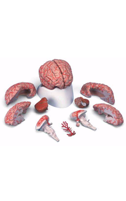
Main Model
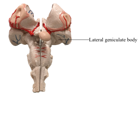
Diencephalon : 30 Lateral geniculate body
Dorsal Thalamus (Thalamus)
The dorsal thalamus (or thalamus) is a massive collection of neuronal cell groups that participate in a widely diverse array of functions involving motor, sensory, and limbic systems. It receives a variety of ascending inputs and projects, via thalamocortical fibers to various cortical areas or gyri, and receives reciprocal connections, via corticothalamic fibers, from those cortical targets to which it sends projections. As a result, the thalamus is often regarded as the functional "gateway" to the cerebral cortex.
The thalamus is covered on its lateral aspect by a layer of myelinated axons, the external medullary lamina, which includes fibers that enter or leave the subcortical white matter. Within the external medullary lamina are clusters of neurons that form the thalamic reticular nucleus. The medial surface of the thalamus borders the third ventricle, and the external medullary lamina and thalamic reticular nucleus blend with the thalamic fasciculus and zona incerta, respectively, to form an interface between dorsal and ventral thalami.
An internal medullary lamina, also consisting of myelinated fibers, extends into the substance of the thalamus, where it forms partitions or boundaries that divide the thalamus into its principal cell groups: the anterior, medial, lateral, and intralaminar nuclear groups. The last cell group is located in the portion of the internal medullary lamina that separates the lateral and medial nuclear groups. In addition, there are midline thalamic nuclei located just superior to the hypothalamic sulcus.
Finally, attached to the caudolateral portion of the thalamus are the medial and lateral geniculate bodies (and their correspondingly named subjacent nuclei). Although considered here as components of the lateral nuclear group, the geniculate nuclei are sometimes considered as a separate part of the thalamus, the metathalamus.
Anterior Thalamic Nuclei
The anterior nucleus forms a prominent wedge on the rostral aspect of the dorsal thalamus just caudolateral to the interventricular foramen; this wedge is the anterior thalamic tubercle. Internal to the anterior tubercle is a large principal nucleus and two smaller nuclei that collectively form the anterior nucleus of the thalamus. Rostrally, the internal medullary lamina divides to partially encapsulate the anterior nucleus. The cells of this nucleus receive dense limbic-related projections from (1) the mammillary nuclei via the mammillothalamic tract and (2) the medial temporal lobe (hippocampus) via the fornix. The output of this nucleus is primarily directed to the cingulate gyrus through the anterior limb of the internal capsule. The anterior nucleus is an important synaptic station in the Papez circuit, which is related to emotion and memory acquisition.
Medial Thalamic Nuclei
This region of the dorsal thalamus comprises the dorsomedial nucleus. This expansive group of neuronal cell bodies is composed of large parvicellular (located caudally) and magnocellular (located rostrally) parts and a small paralaminar part adjacent to the internal medullary lamina. The two larger portions are linked to parts of the frontal and temporal lobes and to the amygdaloid complex. Cells of the paralaminar subdivision receive input from the frontal lobe and substantia nigra and may play a role in the control of eye movement.
Lateral Thalamic Nuclei
This large collection of thalamic neurons is grouped into dorsal and ventral tiers. The relatively small group of dorsal tier nuclei includes the lateral dorsal and lateral posterior nuclei along with the much larger pulvinar nucleus (pulvinar). The connections of the lateral dorsal and lateral posterior nuclei are formed with the cingulate gyrus and parietal lobe, respectively. The large pulvinar nucleus consists of anterior, medial, lateral, and inferior subdivisions. The inferior division receives input from the superior colliculus and projects to the visual association cortex. Other portions of the pulvinar project to areas of the temporal, parietal, and frontal lobes that are especially concerned with visual function and eye movements.
The large ventral tier of the lateral group consists of three separate nuclei. The ventral anterior nucleus (VA) and the slightly more caudal ventral lateral nucleus (VL) are important motor related nuclei; the ventral posterior nucleus, consisting of ventral posterolateral (VPL) and ventral posteromedial (VPM) nuclei, convey somatosensory information to the cerebral cortex.
The VA is composed of a large parvocellular portion and a small magnocellular part. The former receives input from the medial segment of the globus pallidus, and the latter receives afferents from the reticular portion of substantia nigra. The efferent projections from the VA are diffuse and appear to include selected parts of the frontal lobe.
The VL is also composed of three subdivisions: a pars oralis, a pars medialis, and a pars caudalis. The largest of these, the pars oralis, receives a dense projection from the internal segment of the ipsilateral globus pallidus; some of these afferents enter the caudal subdivision. In contrast, the pars caudalis subdivision of the VL receives its main input from the contralateral cerebellar nuclei. Consequently, pallidal and cerebellar projections are largely segregated within this nucleus. The output of the VL reflects its segregated input in that the oral and caudal parts project to largely separate areas of the frontal lobe.
The larger and more laterally located VPL nucleus and the comparatively smaller and more medially located VPM nucleus both receive somatosensory input from the contralateral side of the body. The medial lemniscus and spinothalamic fibers terminate in a somatotopic manner (cervical fibers medial, sacral fibers lateral) within the VPL, whereas trigeminothalamic fibers from the spinal trigeminal nucleus and the principal trigeminal sensory nucleus terminate in the VPM. Both the VPL and VPM project to the somatosensory cortex of the parietal lobe.
A small group of cells called the ventral posterior inferior nucleus is situated ventrally between the VPL and VPM. These cells process vestibular input and project to lateral areas of the postcentral gyrus that are located in the depths of the central sulcus. Similarly, a small group of cells forming the rostral (oral) portion of the VPL receives cerebellar input and projects to the precentral gyrus of the frontal lobe; this nucleus probably represents a few cells that have been displaced from the slightly more rostrally located VL. This cell group is also called the ventral intermediate nucleus because of its location between the VL and VPL.
The medial (MGB) and lateral (LGB) geniculate nuclei are considered parts of the lateral thalamic nuclear group. The MGB receives ascending auditory input via the brachium of the inferior colliculus and projects to the primary auditory cortex in the temporal lobe. The LGB receives visual input from the retina via the optic tract and in turn projects to the primary visual cortex on the medial surface of the occipital lobe.
Located in the posterior thalamus at about the level of the pulvinar and geniculate nuclei is a cluster of cell groups collectively called the posterior nuclear complex. This complex consists of the suprageniculate nucleus, the nucleus limitans, and the posterior nucleus. These nuclei are positioned superior to the medial geniculate and medial to the rostral pulvinar. The posterior nuclear complex receives and sends to the cortex nociceptive cutaneous input that is transmitted over somatosensory pathways.
Intralaminar Nuclei
Embedded within the internal medullary lamina are the discontinuous groups of neurons that form the intralaminar nuclei. These cells are characterized by their projections to the neostriatum and to other thalamic nuclei, along with diffuse projections to the cerebral cortex. Two of the most prominent cell groups are the centromedian and parafascicular nuclei. The centromedian nucleus projects to the neostriatum and to motor areas of the cerebral cortex, whereas the parafascicular nucleus projects to rostral and lateral areas of the frontal lobe. Other intralaminar nuclei receive input from ascending pain pathways and project to the somatosensory and parietal cortex.
Midline Nuclei
The midline nuclei are the least understood components of the thalamus. The largest is the paratenial nucleus, which is located just ventral to the rostral portion of the stria medullaris thalami; other cells are associated with the interthalamic adhesion (massa intermedia). Although inputs are poorly defined, efferent fibers reach the amygdaloid complex and the anterior cingulate cortex, suggesting a role in the limbic system.
Thalamic Reticular Nucleus
The cells of this nucleus are situated within the external medullary lamina and between this lamina and the internal capsule. Axons of these cells project medially into the nuclei of the dorsal thalamus or to other parts of the reticular nucleus, but not into the cerebral cortex. Afferents are received from the cortex and from nuclei of the dorsal thalamus via collaterals of thalamocortical and corticothalamic axons. It appears that thalamic reticular neurons modulate, or gate, the responses of thalamic neurons to incoming cerebral cortical input.