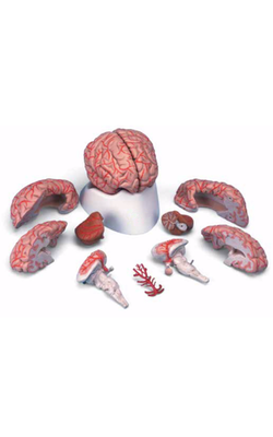
Main Model
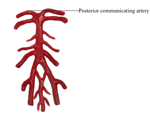
Cerebral arterial circle (Circle of Willis) : 5 Posterior communicating artery
Arterial Blood Supply to Brain
Although it accounts for only about 2.5% of body weight, the brain receives about one sixth of the cardiac output and one fifth of the oxygen consumed by the body at rest. The blood supply to the brain is derived from the internal carotid and vertebral arteries, the terminal branches of which lie in the subarachnoid space. Venous drainage from the brain occurs via cerebral and cerebellar veins that drain to the adjacent dural venous sinuses.
Internal Carotid Arteries
The internal carotid arteries arise in the neck from the common carotid arteries. The cervical part of each artery ascends vertically through the neck, without branching, to the cranial base. Each internal carotid artery enters the cranial cavity through the carotid canal in the petrous part of the temporal bone. In addition to the carotid arteries, the carotid canals contain venous plexuses and carotid plexuses of sympathetic nerves. The internal carotid arteries course anteriorly through the cavernous sinuses, with the abducent nerves (CN VI) and in close proximity to the oculomotor (CN III) and trochlear (CN IV) nerves, running in the carotid groove on the side of the body of the sphenoid. The terminal branches of the internal carotid arteries are the anterior and middle cerebral arteries.
Clinically, the internal carotid arteries and their branches are often referred to as the anterior circulation of the brain. The anterior cerebral arteries are connected by the anterior communicating artery. Near their termination, the internal carotid arteries are joined to the posterior cerebral arteries by the posterior communicating arteries, completing the cerebral arterial circle around the interpeduncular fossa, the deep depression on the inferior surface of the midbrain between the cerebral peduncles.
Vertebral Arteries
The vertebral arteries begin in the root of the neck (the prevertebral parts of the vertebral arteries) as the first branches of the first part of the subclavian arteries. The two vertebral arteries are usually unequal in size, the left being larger than the right. The cervical parts of the vertebral arteries ascend through the transverse foramina of the first six cervical vertebrae. The atlantic parts of the vertebral arteries (parts related to the atlas, vertebra C1) perforate the dura and arachnoid and pass through the foramen magnum. The intracranial parts of the vertebral arteries unite at the caudal border of the pons to form the basilar artery. The vertebrobasilar arterial system and its branches are often referred to clinically as the posterior circulation of the brain.
The basilar artery, so-named because of its close relationship to the cranial base, ascends the clivus, the sloping surface from the dorsum sellae to the foramen magnum, through the pontocerebellar cistern to the superior border of the pons. It ends by dividing into the two posterior cerebral arteries.
Cerebral Arteries
In addition to supplying branches to deeper parts of the brain, the cortical branches of each cerebral artery supply a surface and a pole of the cerebrum. The cortical branches of the:
• Anterior cerebral artery supply most of the medial and superior surfaces of the brain and the frontal pole.
• Middle cerebral artery supply the lateral surface of the brain and the temporal pole.
• Posterior cerebral artery supply the inferior surface of the brain and the occipital pole.
Cerebral Arterial Circle
The cerebral arterial circle (of Willis) is a roughly pentagon-shaped circle of vessels on the ventral surface of the brain. It is an important anastomosis at the base of the brain between four arteries (two vertebral and two internal carotid arteries) that supply the brain.
The arterial circle is formed sequentially in an anterior to posterior direction by the:
• Anterior communicating artery.
• Anterior cerebral arteries.
• Internal carotid arteries.
• Posterior communicating arteries.
• Posterior cerebral arteries.
The various components of the cerebral arterial circle give numerous small branches to the brain.
Venous Drainage of Brain
The thin-walled, valveless veins draining the brain pierce the arachnoid and meningeal layers of dura to end in the nearest dural venous sinuses, which ultimately drain for the most part into the internal jugular veins (IJVs). The superior cerebral veins on the superolateral surface of the brain drain into the superior sagittal sinus; inferior and superficial middle cerebral veins from the inferior, postero-inferior, and deep aspects of the cerebral hemispheres drain into the straight, transverse, and superior petrosal sinuses. The great cerebral vein (of Galen) is a single, midline vein formed inside the brain by the union of two internal cerebral veins; it ends by merging with the inferior sagittal sinus to form the straight sinus. The cerebellum is drained by superior and inferior cerebellar veins, draining the respective aspect of the cerebellum into the transverse and sigmoid sinuses.