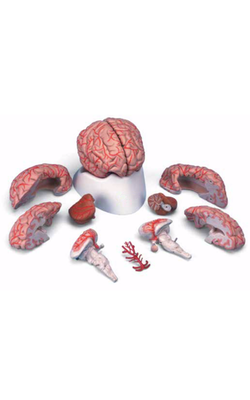
Main Model
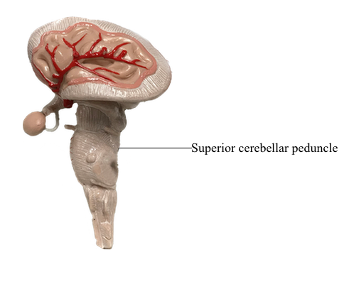
Metencephalon : f Superior cerebellar peduncle
Midpontine Level
Prominent features of the tegmentum at this level include the principal (or chief) sensory trigeminal nucleus, the trigeminal motor nucleus, and the mesencephalic tract and nucleus. The principal sensory and motor trigeminal nuclei are located in the lateral tegmentum, and the mesencephalic tract and nucleus extend rostrally in the lateral wall of the central gray. Cells of the principal sensory nucleus receive SA input from the ipsilateral trigeminal nerve and project to the thalamus via posterior trigeminothalamic (uncrossed) and anterior trigeminothalamic (crossed) fibers. The SE cells of the trigeminal motor nucleus innervate the masticatory muscles on the ipsilateral side. Last, the unipolar cell bodies of the mesencephalic nucleus and their laterally adjacent processes, the mesencephalic tract, convey SA proprioceptive input to a variety of nuclei, including the trigeminal motor nucleus.
The nucleus (locus) ceruleus is located in the lateral floor of the fourth ventricle at this level. Ceruleus neurons contain pigment (hence the alternative name nucleus pigmentosus pontis) and constitute the largest single location of noradrenaline/ norepinephrine-containing cells in the central nervous system. Axons arising in the nucleus ceruleus project to widespread targets throughout the cerebral cortex, most of the diencephalon, limbic system, brainstem, cerebellar cortex and nuclei, and spinal cord. This extensive projection arises from a cell group containing only about 15,000 cell bodies, which indicates that these fibers branch profusely. When the activity level of the neurons in this nucleus is low, a state of quiescence is promoted, as in sleep. When there are sudden changes in the patient’s environment, such as waking unexpectedly or if facing a threatening situation, activity of the nucleus ceruleus is high, noradrenaline/norepinephrine is released throughout the nervous system, and the individual is able to attend to the emergency or disruption. During times of normal nonstress and nonsleep activity, these neurons have an intermediate level of activity. However, when the activity of nucleus ceruleus neurons fluctuates outside of levels that are correlated with normal life activities, the patient may experience behavior problems that require medical or psychiatric intervention.
Most major tracts in the pontine tegmentum (medial longitudinal fasciculus, tectobulbospinal system, medial lemniscus, anterolateral system, and anterior trigeminothalamic fibers) occupy positions comparable to those seen at more caudal levels. Consequently, this section emphasizes only the features that are new at midpontine levels. The medial lemniscus is oriented horizontally at this point, and the rubrospinal fibers have shifted medial to the anterolateral system. Most auditory fibers are now concentrated in the lateral lemniscus, and rostral parts of the superior olivary nucleus appear just lateral to the central tegmental tract. Anterior spinocerebellar fibers migrate posteriorly and enter the cerebellum by coursing over the surface of the superior cerebellar peduncle. The brachium conjunctivum (superior cerebellar peduncle) arises from the cerebellar nuclei, sweeps rostrally, forming the lateral wall of the fourth ventricle, and enters the caudal midbrain tegmentum, where it decussates.
Neurons in the ventral tegmentum close to the midline extend into posterior portions of the basilar pons and constitute the reticulotegmental nucleus. This cell group is continuous with the basilar pontine nuclei, and its axons enter the cerebellum through the contralateral brachium pontis. These cells also share similar features and connections with neurons of the basilar pons.