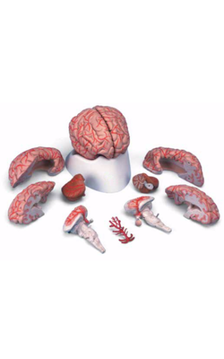
Main Model
Telencephalon : 15 Internal capsule
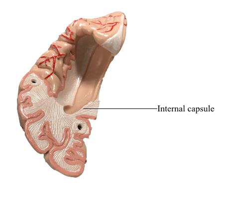
Projection Fibers: The Internal Capsule
The projection fibers of the hemispheres include both the axons that originate outside the telencephalon and project to the cerebral cortex (corticopetal) and the axons that arise from cerebral cortical cells and project to downstream targets (corticofugal). A prime example of the former are projections from the thalamus to the cerebral cortex (thalamocortical fibers); examples of the latter are corticospinal, corticopontine, and corticothalamic fibers. Projection fibers are organized into a large, compact bundle called the internal capsule, which has intimate structural associations with the diencephalon and basal nuclei. Consequently, to divide the internal capsule into its constituent parts, reference must be made to these adjacent cell groups.
In an axial plane through the hemisphere, the internal capsule appears as a prominent V-shaped structure with the V pointing medially. It is divided into three parts: (1) an anterior limb insinuated between the head of the caudate nucleus and the lenticular nucleus, (2) a posterior limb located between the dorsal thalamus and the lenticular nucleus, and (3) a genu located at the intersection of the anterior and posterior limbs, which is located approximately at the level of the interventricular foramen.
The anterior limb of the internal capsule contains thalamocortical-corticothalamic fibers (collectively called the anterior thalamic radiations) that interconnect the dorsomedial and anterior thalamic nuclei with areas of the frontal lobe and the cingulate gyrus. Frontopontine fibers, especially those from the prefrontal areas, also pass through this structure.
The genu of the internal capsule contains corticonuclear fibers that arise in the frontal cortex just rostral to the precentral sulcus and from the precentral gyrus (primary motor cortex) and project to the motor nuclei of cranial nerves. Lesions of these fibers give rise to motor deficits of cranial nerves, most notably deficits related to the facial and hypoglossal nerves.
The posterior limb of the internal capsule is larger and more complex. It is sometimes divided into a thalamolenticular part (located between the thalamus and the lenticular nucleus), a sublenticular part (fibers passing ventral to the lenticular nucleus), and a retrolenticular part (fibers located caudal to the lenticular nucleus). However, contemporary terminology and common usage refer to the thalamolenticular part as the posterior limb, the sublenticular part as the sublenticular limb, and the retrolenticular part as the retrolenticular limb. This terminology is considerably less cumbersome and much easier to remember and is the convention followed here. By this scheme, the internal capsule consists of five parts: anterior limb, genu, posterior limb, sublenticular limb, and retrolenticular limb.
Included in the posterior limb are corticospinal fibers arising from the motor cortex and projecting to the contralateral spinal cord and thalamocortical-corticothalamic fibers (as part of the central thalamic radiations) that interconnect nuclei of the dorsal thalamus with the overlying cortex. Studies in humans have revealed that corticospinal fibers are somatotopically arranged in about the caudal half of the posterior limb. Geniculotemporal radiations (auditory radiations) convey auditory information from the medial geniculate nucleus to the transverse temporal gyri through the sublenticular limb. Visual input from the lateral geniculate body to the occipital cortex is conveyed via geniculocalcarine radiations (optic radiations) through the retrolenticular limb. Optic radiations form a distinct lamina of fibers immediately lateral to the tapetum as they course caudally into the occipital lobe.
Fibers of the internal capsule flare out into the hemisphere as they pass distal to the caudate and putamen. This abrupt divergence of internal capsule fibers forms the corona radiata (“radiating crown”), which contains converging corticofugal fibers as well as diverging corticopetal fibers.
Vasculature of the Internal Capsule
The blood supply to the genu and most of the posterior limb of the internal capsule is via the lenticulostriate arteries; these are branches of the M1 segment. Branches of the anterior choroidal artery supply the inferior region of the posterior limb, optic tract, portions of the optic radiation (the Meyer loop), inferior parts of the basal nuclei, hippocampus, amygdala, choroid plexus in the temporal horns, and immediately adjacent retrolenticular limb. An occlusion of this vessel gives rise to a constellation of deficits, called the anterior choroidal artery syndrome, reflecting damage to these structures.
The anterior limb receives somewhat of a dual blood supply in that lenticulostriate arteries and branches of the medial striate artery (usually a branch of A2) serve this area.
Whereas a variety of clinical events may damage the fibers of the internal capsule, the vast majority (about 95%) that produce rapid-onset deficits are vascular related. Hemorrhagic stroke (about 15% of cases: rupture of or bleeding from a vessel serving the capsule) and occlusive stroke (about 85% of cases: occlusion of a vessel serving the capsule) are the most common causes. Lesions of the posterior limb may result in a combination of motor (corticospinal tract involvement) and sensory (thalamocortical fiber involvement) deficits that are seen on the side of the body contralateral to the lesion. Indeed, a characteristic feature of hemisphere lesions is the appearance of motor and sensory deficits on the same side of the body. Lesions of the retrolenticular limb result in visual deficits (optic radiation fiber involvement) that may, depending on the extent of the damage, involve the contralateral hemifield (one half of each visual field, a hemianopia) or a contralateral quadrant (about one fourth of each visual field, a quadrantanopia) of the visual field of each eye. Damage to the sublenticular limb may result in tinnitus and difficulty localizing sound.