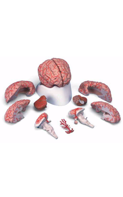
Main Model
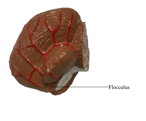
Metencephalon : c Flocculus
Cerebellar Lobes, Lobules, and Zones
At the most general level, it is common to divide the cerebellum
into a narrow midline vermis and expansive lateral hemispheres. The cerebellum is further
divided into anterior, posterior, and flocculonodular lobes by the
primary and posterolateral fissures, respectively. The lobes of the
cerebellum are composed of yet smaller divisions called lobules. The lobules of the vermis are
identified by Roman numerals I to X; the corresponding lateral
(hemisphere) portion of each vermis lobule is identified by the same
Roman numeral but with the prefix H. Vermis lobules II to X have hemisphere portions HII to HX;
lobule I does not have a hemisphere part in humans. The anterior
lobe comprises lobules I to V and HII to HV, and the posterior
lobe, lobules VI to IX and HVI to HIX. The flocculonodular lobe
consists of the nodulus (lobule X) and its hemisphere counterpart,
the flocculus (lobule HX). In turn, each lobule consists of a series of
individual ridges of cortex called folia (singular, folium).
The individual folia are continuous from one hemisphere to
the other, across the midline on the superior cerebellar surface. This pattern, obvious on the superior cerebellar surface, is disrupted on the inferior surface by the enlargement
of the lateral parts of the cerebellum and consequent infolding of
the midline area.
Superimposed on the lobes and lobules of the cerebellum
are rostrocaudally oriented cortical zones that are defined on
the basis of their connections. There are three principal zones
on each side: the medial (vermal), intermediate (paravermal),
and lateral (hemisphere) zones. On the basis of their
afferent and efferent connections, these three larger cortical
zones can be subdivided further into nine smaller zones. In general, these zone patterns are the basis for the modules. The clinical deficits that result from a cerebellar lesion depend mainly on which of the three principal zones
is involved; consequently, the three-zone terminology is used.
The medial (vermal) zone is a narrow strip of cortex adjacent to the midline that extends throughout anterior and posterior lobes and includes the nodulus.
This zone is widest in lobule VI and tapers rostrally and caudally. The intermediate (paravermal) zone lies adjacent to the medial zone and extends throughout anterior and posterior
lobes but has little representation in the flocculonodular lobe. The lateral (hemisphere) zone occupies by far the
largest part of the cerebellar cortex. It includes large portions
of anterior and posterior lobes and the flocculus.