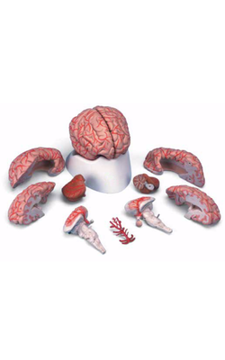
Main Model
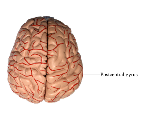
Telencephalon : 7 Postcentral gyrus
Parietal Lobe
The parietal lobe consists of the postcentral gyrus, located between the central and postcentral sulci, and the superior and inferior parietal lobules, which are separated by the intraparietal sulcus. Gyri forming the superior parietal lobule extend onto the medial surface of the hemisphere as the precuneus, whereas the inferior parietal lobule is made up of the angular and supramarginal gyri. The latter is a crescent-shaped ridge of cortex around the caudal terminus of the lateral sulcus.
As the postcentral gyrus extends onto the medial surface of the hemisphere, it is continuous with the posterior paracentral gyrus. This cortical area is bordered rostrally by an imaginary line that connects the central sulcus to the cingulate sulcus and caudally by the marginal ramus of the cingulate sulcus. The latter is frequently called the marginal sulcus. Taken together, the postcentral gyrus and posterior paracentral lobule constitute the primary somatosensory cortex.
Specific functional areas in the parietal lobe include the primary somatosensory cortex (Brodmann areas 3, 1, 2) and the gyri that are part of the Wernicke area (supramarginal gyrus - Brodmann area 40 and the angular gyrus - Brodmann area 39). Clinically, the Wernicke area is believed to extend into the temporal lobe and to encompass portions of Brodmann area 22 and some of area 21. The body is somatotopically organized in the postcentral and posterior paracentral gyri in a pattern generally similar to that seen in the precentral gyrus. Within the postcentral gyrus, the face is represented in the lateral third, the upper extremity (with emphasis on the fingers) in the middle third, and the trunk, hip, and thigh in about the medial third; the leg, foot, and genitalia are represented in the posterior paracentral lobule. Damage to the primary somatosensory cortex results in an alteration of sensory (pain, thermal, and proprioception) perception.
The angular gyrus (Brodmann area 39) and the supramarginal gyrus (Brodmann area 40) collectively form a portion of the Wernicke area; these two gyri also comprise the inferior parietal lobule. Lesions of the Wernicke area result in a constellation of deficits called Wernicke aphasia (or receptive aphasia). These patients cannot understand what they hear, cannot read or write, and speak in a jumble of words that makes no sense. Theirs is a receptive problem; information is received but it cannot be understood or used to express coherent thought.