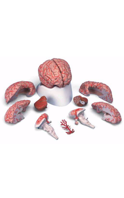
Main Model
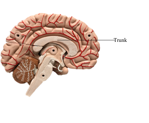
Telencephalon : b Trunk
Commissural Fibers: The Corpus Callosum
In general, commissural fibers interconnect corresponding structures on either side of the neuraxis. The largest bundle of commissural fibers is the corpus callosum. This huge bundle is located superior to the diencephalon and forms the roof of much of the lateral ventricles. The corpus callosum consists of, from rostral to caudal, a rostrum, genu, body (also called trunk), and splenium. Many of the fibers passing through the genu arch rostrally to interconnect the frontal lobes; these form the minor (or frontal) forceps. The fibers interconnecting the occipital lobes loop through the splenium of the corpus callosum, forming the major (or occipital) forceps. The tapetum, which is located in the lateral wall of the atrium and posterior horn of the lateral ventricle, is also composed of fiber bundles that cross in the splenium.
Lesions of the corpus callosum, whether the result of surgery or spontaneous, disconnect one hemisphere from the other. About 80% of patients who have severe generalized seizures experience significant relief after section of the anterior 75% of the corpus callosum, largely sparing the splenium. Spontaneous lesions of the corpus callosum may result from vascular infarct, tumor (such as oligodendroglioma), or necrosis or demyelination (as in Marchiafava-Bignami disease). It is recognized that some of these patients may have a disconnection syndrome.
Smaller commissural bundles include the anterior commissure and the hippocampal commissure. In sagittal views, the anterior commissure is located caudal to the rostrum of the corpus callosum but rostral to the main part of the fornix. This bundle interconnects various parts of the frontal and temporal lobes. The hippocampal commissure is formed by fibers that originate in the hippocampal formations and cross the midline as a thin layer inferior to the splenium of the corpus callosum.
The posterior commissure and the habenular commissure are small fiber bundles traversing the midline that connect caudal parts of the diencephalon. The former crosses the midline at the base of the pineal gland and just posterior (dorsal) to the cerebral aqueduct. The latter is a small fascicle running along the upper aspect of the posterior commissure and interconnecting the habenular nuclei.