Main Model
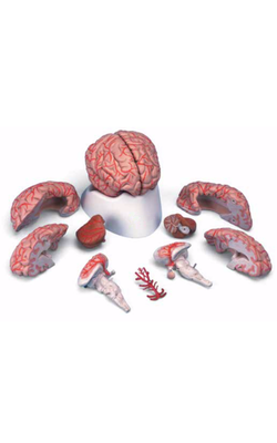
Telencephalon : View 1
The telencephalon is the largest part of the human brain, constituting about 85% of total brain weight, and is that portion in which all modalities are represented. Various sensory inputs (such as vision and hearing) are localized in some areas, whereas motor functions are represented in other regions and are modulated by subcortical nuclei. The telencephalon contains circuits that interrelate regions that have specific functions, such as motor or visual, with other regions called association areas. Seeing a familiar image may precipitate a cascade of neural events having olfactory, emotional, sensory, and motor components. Damage to association areas results in complex neurologic deficits. The patient may not be blind or paralyzed but may be unable to recognize sensory input (agnosia), express ideas or thoughts (aphasia), or perform complex goal-directed movements (apraxia).
Overview
The telencephalon consists of large hemispheres separated from each other by a deep longitudinal cerebral fissure. Each hemisphere has an outer surface, the cerebral cortex, which is composed of layers of cells. The cortex is thrown into elevations called gyri (singular, gyrus) that are separated by grooves called sulci (singular, sulcus). Internal to the cortex are large amounts of subcortical white matter along with aggregates of gray matter that form the basal nuclei and the amygdala. Although not parts of either the telencephalon or the basal nuclei, the subthalamic nucleus (of the diencephalon) and the substantia nigra (of the mesencephalon) have important connections that functionally link them with the basal nuclei.
Information passing into or out of the cerebral cortex must traverse the subcortical white matter. The myelinated fibers forming the white matter are organized into (1) association bundles that connect adjacent or distant gyri in one hemisphere; (2) commissural bundles that connect the hemispheres, the largest of these being the corpus callosum; and (3) the internal capsule. The internal capsule contains axons projecting to numerous downstream nuclei (corticofugal fibers) and axons conveying information to the cerebral cortex (corticopetal fibers). The terms corticofugal and corticopetal are umbrella terms that include all efferent and all afferent fibers, respectively, of the cerebral cortex.
The hippocampal complex and the amygdala are located in the walls of the temporal horn of the lateral ventricle. The axons of cells in these structures coalesce to form the fornix, stria terminalis, and amygdalofugal pathway.
Development
Enlargements of the prosencephalon, the telencephalic (cerebral) vesicles, appear at about 5 weeks of gestation. As the cerebral vesicles enlarge in all directions, they pull along portions of the neural canal that will form the cavities of the telencephalon, the lateral ventricles. The primitive lateral ventricles extend into frontal, parietal, temporal, and occipital areas as they develop and form that portion of the ventricle found in each of these lobes in the adult. The interventricular foramina, which connect each lateral ventricle to the midline third ventricle (cavity of the diencephalon), are initially large but become smaller as development progresses. In the adult brain, each interventricular foramen is bordered rostromedially by the column of the fornix and caudolaterally by the anterior tubercle of the dorsal thalamus.
Cells forming the corpus striatum appear in the floor of the developing lateral ventricle at the time when primordial cell groups in the wall of the third ventricle are giving rise to diencephalic structures. As development progresses, the corpus striatum is bisected by axons growing to and from the cerebral cortex. These axons form the internal capsule of the adult and divide the corpus striatum into a medially located caudate nucleus and a laterally located putamen. As the diencephalon enlarges, it gives rise to the thalamus and hypothalamus and to cells that migrate across the developing internal capsule to assume a position medial to the putamen. These cells become the globus pallidus of the adult and, in combination with the putamen, form the lenticular nucleus.
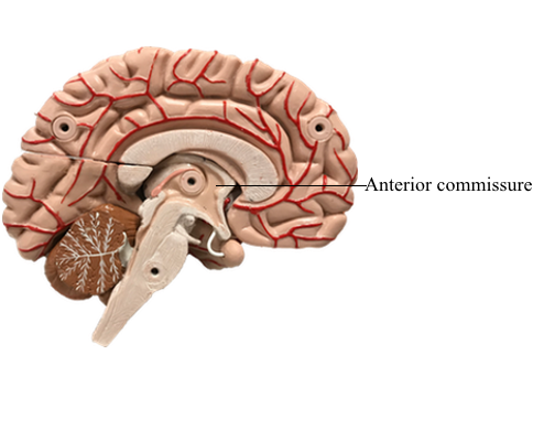
The initial development of the major commissural bundles and of the hippocampus takes place along the medial aspect of the hemisphere. In the adult brain, there are three major interhemispheric commissures: the anterior commissure, the hippocampal commissure, and the corpus callosum. The first of these to appear, the anterior commissure, arises within the lamina terminalis, a membrane-like structure that extends from the anterior commissure anteriorly (ventrally) to the rostral edge of the optic chiasm. The second to form, the hippocampal commissure, develops along with the hippocampal primordium. As growth occurs, the hippocampus, which originates in the posteromedial part of the hemisphere, is displaced into the temporal lobe, where it assumes a position characteristic of the adult. In the process, fibers from one side cross to the other side, as the hippocampal commissure, just inferior to the area that will be occupied by the corpus callosum. The third commissure to develop, the corpus callosum, originates from the area of the lamina terminalis as a structure initially composed of astrocytic processes. Axons from developing neurons in each hemisphere traverse this glial structure to access the contralateral side. As this takes place, the corpus callosum enlarges in a caudal direction to form the prominent structure found in the adult.
Developmental Defects
There are numerous developmental events that may cause defects in the configuration of the telencephalon. One of the developmental failures that will result in aberrant development of the telencephalon is the improper migration of maturing neurons on radial glia. This failure results in structural and in some cases corresponding functional defects in the arrangement of the cerebral cortex. Some examples include lissencephaly (a lack of gyri and sulci, a smooth brain), pachygyria (abnormally large gyri that are few in number), and microgyria (abnormally small gyri that are greater in number).
Holoprosencephaly is a preneurulation defect that is represented by three general forms. Alobar holoprosencephaly, the most severe form, consists of a midline ventricle, no hemispheres or corpus callosum, and severe retardation. Semilobar holoprosencephaly consists of a partial formation of lobes with the ventricles formed; the frontal lobes may be fused, and the occipital lobes may be separated by an incomplete longitudinal fissure. Although the ventricles are formed, midline structures such as the septum pellucidum are missing. In lobar holoprosencephaly, the least severe form, the longitudinal fissure is largely complete, hemispheres exist, generally normal patterns of sulci and gyri are seen, but there is a fusion of the hemispheres at the frontal pole or at the orbital surface of the frontal lobe.
Anencephaly is a severe developmental failure in which the telencephalon and the surrounding skull are largely absent. This defect is catastrophic and not compatible with life. Anencephaly is generally associated with a failure of the anterior neuropore to close. The lamina terminalis represents the adult position of the anterior neuropore.
Failure of the corpus callosum to develop (agenesis of the corpus callosum) may be accompanied by an absence of the anterior and hippocampal commissures. Although some patients with this condition may experience focal seizures and have mental retardation, others live for many years with few or no obvious neurologic deficits. These individuals frequently have developmental abnormalities in other parts of the nervous system.