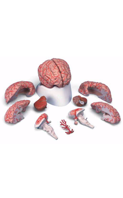
Main Model
Metencephalon : a Vermis
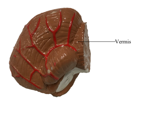
Cerebellum
The cerebellum is located posterior to the brainstem and fills much of the posterior fossa. It is attached to the brainstem by three pairs of cerebellar peduncles (superior, middle, and inferior). In sagittal section, the human cerebellum appears wedge shaped, with its superior surface apposed to the tentorium cerebelli and its inferior surface curving toward the foramen magnum.
The cerebellum consists of anterior, posterior, and flocculonodular lobes; each lobe, in turn, is composed of lobules. Lobes and lobules are separated from each other by fissures. Lobules are made up of yet smaller folds of cerebellar cortex called folia (singular, folium). Cerebellar folia, lobules, and lobes can often be followed across the midline from one side of the cerebellum to the other.
Each cerebellar lobe (and lobule) is also divided into rostrocaudally oriented regions of cortex commonly called the vermis (medial), intermediate (paravermis), and hemisphere (lateral) zones. The vermal zone is approximately 1.0 cm across at its widest point. The hemisphere is expansive in the human cerebellum and is separated from the vermis by the intermediate zone.
Four cerebellar nuclei are located in the white matter core of each hemisphere. From medial to lateral, they are the fastigial, globose, emboliform, and dentate nuclei. These cells receive input from branches of cerebellar afferent fibers and from Purkinje cells located in the cerebellar cortex. In turn, axons of cerebellar nuclear cells provide the main output signals of the cerebellum.