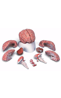
Main Model
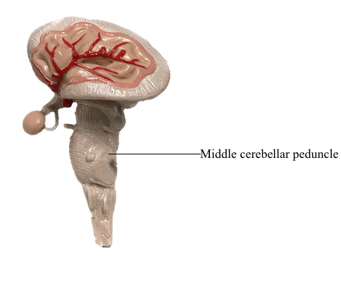
Metencephalon : g Middle cerebellar peduncle
Caudal Pontine Level
The pons is divided into a posterior part, the tegmentum, and an anterior region, the basilar pons. This section and the following two sections describe the anatomy of the pons at three levels: caudal, midpontine, and rostral pontine levels. Each level is described beginning with the tegmentum and proceeding to the basilar pons.
At caudal pontine levels, the facial colliculus is formed by the underlying abducens nucleus and fibers comprising the internal genu of the facial nerve. Axons from the SE cells of the abducens nucleus course anteriorly through the tegmentum, pass adjacent to the corticospinal fibers in the basilar pons, and exit the brainstem at the pons-medullary junction as the abducens nerve. The internal genu of cranial nerve VII is composed of the axons of SE cells from the facial nucleus. These axons loop around the abducens nucleus from caudal to rostral, as the internal genu, then course anterolaterally to exit the brainstem. Anterolateral to the abducens nucleus, these SE fibers are surrounded by cells of the superior salivatory nucleus, the axons of which exit the brainstem as the VE component of the intermediate nerve.
Medial to the abducens nucleus is the medial longitudinal fasciculus and the tectobulbospinal system. As in the medulla, these bundles are internal to the ventricular space and adjacent to the midline.
The posterolateral tegmentum contains the vestibular nuclei and the solitary tract and nucleus. The lateral, medial, and inferior vestibular nuclei are present at this level, whereas the superior vestibular nucleus becomes prominent more rostrally. The small bundles of fibers coursing between the vestibular nuclei and the cerebellum in the wall of the fourth ventricle form the juxtarestiform body. This structure is composed of vestibulocerebellar and cerebellovestibular fibers and along with the laterally adjacent restiform body constitutes the inferior cerebellar peduncle. The rostral portions of the solitary tract and nucleus are located anterior to the vestibular nuclei and consist of a core of primary sensory fibers (tract) surrounded by cell bodies (nucleus). This part of the solitary complex receives mainly taste input (VA fibers) and is sometimes called the gustatory nucleus.
The central portion of the pontine tegmentum at caudal levels contains, from medial to lateral, the central tegmental tract, the superior olivary nucleus, the facial motor nucleus, and the spinal trigeminal tract and nucleus. A major part of the central tegmental tract includes fibers coursing from the red nucleus of the midbrain to the inferior olive of the medulla (rubroolivary fibers). Cells of the superior olive receive input from the anterior cochlear nucleus and send their axons into the lateral lemniscus on both sides. The route is followed by motor facial fibers; these axons innervate the ipsilateral muscles of facial expression. The spinal trigeminal tract is composed of sensory (SA) fibers from the ipsilateral half of the face, oral cavity, and much of the scalp. Although most of this input is via the trigeminal nerve (hence the name of the tract), cranial nerves VII, IX, and X also make modest contributions to the spinal trigeminal tract and nucleus. Axons of the spinal trigeminal tract synapse in the spinal trigeminal nucleus, the cells of which project to the contralateral thalamus as anterior trigeminothalamic fibers.
The anterolateral system, rubrospinal tract, and trapezoid body are located in the anterolateral tegmentum. Although pain and temperature signals from the contralateral side of the body are conveyed by anterolateral system fibers, some of these axons end in the pontine reticular formation as spinoreticular fibers. The trapezoid body is composed of decussating axons from the cochlear nuclei. After crossing, these fibers ascend to form the lateral lemniscus and convey auditory signals to the midbrain.
The medial lemniscus is oriented vertically in the medulla, but in the caudal pons it begins to shift to a horizontal position. At this level, the anterior part of the medial lemniscus (lumbosacral representation) shifts somewhat laterally, and its posterior portion (cervicothoracic representation) assumes a more medial location. The medial lemniscus forms the border between the tegmentum and basilar pons.
The anterior pons contains the basilar pontine nuclei, longitudinally running corticospinal and corticopontine fibers, and transversely oriented pontocerebellar fibers. On each side of the midline, the corticospinal fibers located in the pyramid of the medulla are, in the basilar pons, completely surrounded by the basilar pontine nuclei. These pontine cells receive input from diverse regions of the neuraxis. In turn, most of their axons cross the midline and enter the cerebellum via the middle cerebellar peduncle (brachium pontis) as pontocerebellar fibers.