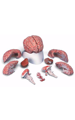
Main Model
Cranial Nerves : V Trigeminal nerve
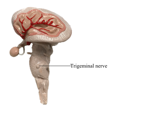
Trigeminal Nerve (CN V)
Functions: Somatic (general) sensory and somatic (branchial) motor to derivatives of the 1st pharyngeal arch.
Nuclei: There are four trigeminal nuclei - one motor (motor nucleus of trigeminal nerve), and three sensory (mesencephalic, principal sensory and spinal nuclei of trigeminal nerve).
The trigeminal nerve (CN V) is the largest cranial nerve (if the atypical optic nerve is excluded). It emerges from the lateral aspect of the pons by a large sensory root and a small motor root. The roots of CN V are comparable to the posterior and anterior roots of spinal nerves. CN V is the principal somatic (general) sensory nerve for the head (face, teeth, mouth, nasal cavity, and dura mater of the cranial cavity). The large sensory root of CN V is composed mainly of the central processes of the pseudounipolar neurons that make up the sensory trigeminal ganglion. The ganglion is flattened and crescent shaped (hence its unofficial name, semilunar ganglion), and is housed within a dural recess (trigeminal cave) lateral to the cavernous sinus.
The peripheral processes of the ganglionic neurons form three nerves or divisions: ophthalmic nerve (CN V1), maxillary nerve (CN V2), and sensory component of the mandibular nerve (CN V3). Maps of the zones of cutaneous innervation by the three divisions resemble the dermatome maps for cutaneous innervation by spinal nerves. Unlike spinal nerve dermatomes, however, there is little overlap in innervation by the divisions; lesions of a single nerve result in clearly demarcated areas of numbness.
The fibers of the motor root of CN V pass inferior to the trigeminal ganglion along the floor of the trigeminal cave, bypassing the ganglion (just as the anterior roots of spinal nerves bypass the spinal sensory ganglia). They are distributed exclusively via the mandibular nerve (CN V3), blending with the sensory fibers as the nerve traverses the foramen ovale in the cranium. Branches pass to the muscles of mastication, mylohyoid, anterior belly of the digastric, tensor veli palatini, and tensor tympani, which are derived from the 1st pharyngeal arch.
Although CN V conveys no presynaptic parasympathetic fibers from the CNS, all four parasympathetic ganglia are associated with the divisions of CN V. Postsynaptic parasympathetic fibers from the ganglia join branches of CN V and are carried to their destinations along with the CN V sensory and motor fibers.
Ophthalmic Nerve (CN V1)
In contrast to the other two CN V divisions, CN V1 is not a branchial nerve (i.e., it does not supply pharyngeal arch derivatives). It serves structures derived from the paraxial mesoderm of the embryonic frontonasal process. The ophthalmic nerve's association with the other CN V divisions is a secondary occurrence. The somatic (general) sensory fibers of CN V1 are distributed to skin and mucous membranes and conjunctiva of the front of the head and nose.
Testing CN V1: The integrity of this division is tested by checking the corneal reflex - touching the cornea, which is also supplied by CN V1, with a wisp of cotton will evoke a reflexive blink if the nerve is functional.
Maxillary Nerve (CN V2)
CN V2 innervates derivatives of the maxillary prominence of the 1st pharyngeal arch. Exiting the cranial cavity via the foramen rotundum, its somatic (general) sensory fibers are generally distributed to skin and mucous membranes associated with the upper jaw. The pterygopalatine (parasympathetic) ganglion is associated with this division of CN V, involved in innervating the lacrimal gland and glands of the nose and palate.
Mandibular Nerve (CN V3)
CN V3 innervates derivatives of the mandibular prominence of the 1st pharyngeal arch. CN V3 is the only division of CN V to convey somatic (branchial) motor fibers, distributed to the striated muscle derived from mandibular prominence mesoderm, primarily the muscles of mastication. Two parasympathetic ganglia, the otic and submandibular, are associated with this division of CN V; both are concerned with the innervation of salivary glands.