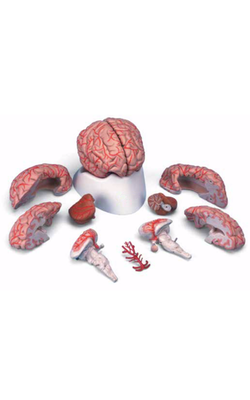
Main Model
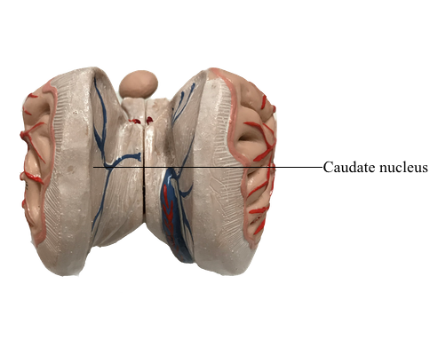
Diencephalon : 27 Caudate nucleus
Caudate and Lenticular Nuclei
The caudate and lenticular nuclei collectively form the corpus striatum. The corpus striatum, in turn, is divided into the neostriatum, consisting of the caudate nucleus and the putamen, and the paleostriatum, or globus pallidus. The globus pallidus and putamen collectively form the lenticular nucleus.
The caudate nucleus is characteristically located in the lateral wall of the lateral ventricle and consists of three parts: head, body, and tail. The head of the caudate nucleus forms a prominent bulge in the anterior horn of the lateral ventricle. In Huntington disease (also called Huntington chorea), an inherited neurodegenerative disease, the head of the caudate nucleus is characteristically diminished in size or absent in magnetic resonance imaging or computed tomography. At about the level of the interventricular foramen, the caudate diminishes in size but continues caudally as the body of the caudate nucleus in the lateral wall of the body of the lateral ventricle. In the lateral wall of the atrium of the lateral ventricle, the body of the caudate nucleus turns inferiorly and rostrally to continue as the tail of the caudate nucleus in the posterolateral (dorsolateral) wall of the temporal horn of the lateral ventricle. Thus the C shape of the caudate nucleus faithfully follows the C shape of the lateral ventricle (excluding the posterior horn).
The lenticular nucleus is located within the base of the hemisphere and is surrounded by white matter. The internal capsule borders the lenticular nucleus medially, and the external capsule separates it from the claustrum laterally. The lenticular nucleus consists of a larger lateral part, the putamen, and a small medial portion, the globus pallidus (or pallidum). The putamen extends more superior, more rostral, and more caudal compared with the globus pallidus and is clearly the larger part of the lenticular nucleus when viewed in axial or coronal planes. The globus pallidus is located internal to the putamen and is smaller in all dimensions. It is divided into medial (internal) and lateral (external) parts by thin sheets of vertically oriented white matter. The globus pallidus is also separated from the putamen by a thin lamina of white matter.