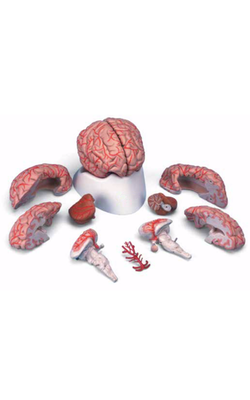
Main Model
Telencephalon : 17 Thalamostriatic vein
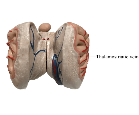
Internal Veins of the Hemisphere
The main venous channels draining internal structures of the hemisphere are the internal cerebral veins. They
course along the dorsomedial edge of the thalamus and are located
in the tela choroidea of the third ventricle. One of the principal tributaries of the internal cerebral veins, the thalamostriate
vein (also called the terminal vein), is found
in association with the stria terminalis and drains the caudate nucleus (via the transverse caudate veins) and internal regions of
the hemisphere dorsal and lateral to the caudate nucleus.
The two internal cerebral veins join to form the great cerebral
vein (of Galen). This large venous channel has several tributaries and is caudally continuous with the straight
sinus.
Cerebral and spinal veins and dural sinuses lack valves. Consequently, pathologic processes may alter normal venous flow
patterns and result in the transport of material into the brain.
For example, a tumor or infection in the orbit may cause venous
blood to flow toward the cavernous sinus rather than away from it. In this way, infectious material or tumor cells may pass from
the orbit into the cavernous sinus and, through its connecting
channels, to other parts of the brain.
Malformations of the great cerebral vein of Galen are sometimes described as a special type of AVM or as an aneurysm. This
lesion is usually seen in newborns or infants. In these
cases, the great cerebral vein is grossly enlarged and fed by large
and abnormal branches of the cerebral and cerebellar arteries.
Bulging fontanelles, progressive hydrocephalus (resulting from occlusion of the cerebral aqueduct), and dilated veins in the face
and scalp are characteristic findings.