Main Model

Brain : III Mesencephalon
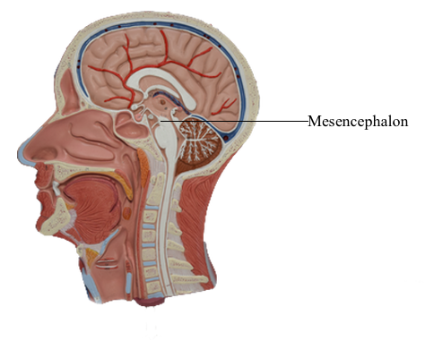
The mesencephalon, or midbrain, is the most rostral portion of
the brainstem. It gives rise to cranial nerves III and IV, conducts
ascending and descending tracts, and contains nuclei that are
essential to motor function. Caudally the midbrain is continuous with the pons, and rostrally it joins the diencephalon. The
cerebral aqueduct, the cavity of the midbrain, is continuous rostrally with the third ventricle and caudally with the fourth ventricle. The blood supply to the mesencephalon is primarily from
proximal branches of the posterior cerebral arteries (P1 or P2)
and from penetrating branches of the posterior communicating
artery.
External Features
Anterior (Ventral) Midbrain
The presence of a pair of large axon bundles, the crura cerebri,
is a characteristic feature of the anterior aspect of the midbrain.
These bundles emerge from the cerebral hemispheres caudal to
the optic tracts, converge slightly toward the midline as they
course through the midbrain, and disappear into the basilar pons. The oculomotor nerves exit the medial edge of each
crus and pass through the space between the crura: the interpeduncular fossa. Anteriorly, the rostral limit of the
midbrain is marked by the exit of the crura cerebri from the
cerebral hemispheres and by the caudal edge of the mammillary
bodies. The caudal border of the midbrain is formed where each
crus enters the basilar pons.
The subarachnoid space of the interpeduncular fossa is called
the interpeduncular cistern. This cistern contains the oculomotor nerves and the upper part of the basilar artery, including its bifurcation and proximal branches. Numerous vessels penetrate
the roof of this fossa and create many small perforations. This area is frequently called the posterior perforated substance.
Posterior (Dorsal) Midbrain
The posterior surface of the adult midbrain is characterized by
four elevations collectively called the corpora quadrigemina. The rostral two elevations are the superior colliculi,
and the caudal two are the inferior colliculi. Just caudal to the
inferior colliculus, the exit of the trochlear nerve marks the pons-midbrain junction on the posterior surface of the brainstem,
whereas the midbrain-diencephalic boundary is formed by the
posterior commissure.
Rostrolaterally, the inferior colliculus is joined to the medial
geniculate body of the diencephalon by a fiber bundle called the
brachium of the inferior colliculus. The inferior colliculus and the medial geniculate body are part of the auditory
system. The brachium of the superior colliculus extends from the
optic tract to the superior colliculus in a groove located between
the medial geniculate body and pulvinar of the diencephalon. The superior colliculus, pulvinar, and lateral
geniculate body are parts of the visual and visual-motor systems.
On the midline, the pineal gland, a diencephalic structure,
extends posteriorly above and between the superior colliculi. Tumors of the pineal may produce noncommunicating (obstructive) hydrocephalus because of compression of the colliculi
of the midbrain and resulting occlusion of the cerebral aqueduct.
The subarachnoid space immediately posterior (dorsal) to
the colliculi is the quadrigeminal cistern. This cistern contains
the exiting trochlear nerves, the great vein of Galen, and distal branches of the posterior cerebral arteries. The ambient cistern
is located at the lateral aspect of the midbrain and contains segments P2 to P3 and the superior cerebellar and quadrigeminal arteries. The crural cistern is located between the
crus cerebri and the immediately adjacent parahippocampal
gyrus; it contains medial posterior choroidal and anterior choroidal arteries and the basal vein (of Rosenthal). The interpeduncular cistern is that part of the subarachnoid space that occupies
the interpeduncular fossa.
Vasculature of the Midbrain
The primary blood supply to the mesencephalon arises via branches
of the basilar artery, with smaller branches from the superior cerebellar, anterior choroidal, medial posterior choroidal, and posterior communicating arteries. An important source of blood to the posterior portion of the midbrain is the quadrigeminal artery,
a branch of the posterior cerebral artery (P1 segment). Also, the
superior cerebellar artery gives rise to branches that serve caudal
parts of the posterior midbrain and adjacent regions of the pons.