Main Model

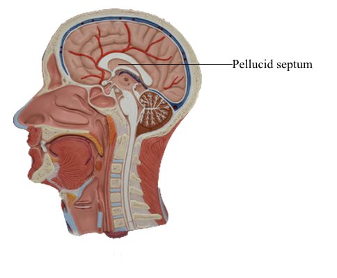
Brain : 5 Pellucid septum
Ventricles
Lateral Ventricles
The cavities of the telencephalon are the lateral ventricles, of
which there is one in each hemisphere. As the development of the hemispheres creates the frontal, temporal, and occipital lobes, the lateral ventricles are pulled along and thus
acquire their definitive adult shape of a flattened C with a short
tail. This shape is present by birth. The lateral ventricle consists of an anterior horn, a body, and posterior and inferior
horns. The junction of the body with the posterior and
inferior horns constitutes the atrium of the lateral ventricle. An especially large clump of choroid plexus, the glomus (or glomus
choroideum), is found in the atrium. In adults and
especially in elderly persons, the glomus may contain calcifications that are visible (as white spots) on radiographs or computed
tomography (CT) scans. Shifts in the position of the
glomus, usually accompanied by alterations in the volume or
shape of the surrounding ventricle, may indicate some type of
ongoing pathologic process or space occupying lesion.
The elaborate shape of the lateral ventricle means that different structures border on different parts of this space. The
anterior horn and body of the lateral ventricle are bordered
medially by the septum pellucidum (at rostral levels) and by a
bundle of fibers called the fornix (at caudal levels) and posteriorly (superiorly) by the corpus callosum.
The floor of the body of the lateral ventricle is made up of the
thalamus, and the caudate nucleus is characteristically found in the lateral wall of the lateral ventricle throughout its extent. In the temporal lobe, the inferior horn of
the lateral ventricle contains the tail of the caudate nucleus in
its lateral wall, the hippocampal formation in its medial wall,
and a large group of cells (the amygdaloid complex) in its rostral end.
The openings between the lateral and third ventricles, the
interventricular foramina, are located between the column of
the fornix and the rostral and medial ends of the thalamus. There
are two interventricular foramina, one opening from each lateral
ventricle into the single midline third ventricle.
Third Ventricle
The third ventricle, the cavity of the diencephalon, is a narrow,
vertically oriented midline space that communicates rostrally
with the lateral ventricles and caudally with the cerebral aqueduct. The third ventricle has an elaborate profile on a sagittal view, but it is narrow in the coronal and axial planes.
The boundaries of the third ventricle are formed by a variety
of structures, the most important being the dorsal thalamus
and hypothalamus, and by structures that form small outpocketings called recesses. These are the supraoptic recess (above the optic chiasm), the infundibular recess
(in the infundibulum, the stalk of the pituitary), the pineal
recess (in the stalk of the pineal), and the suprapineal recess
(above the pineal). The rostral wall of the third ventricle is formed by a short segment of the anterior commissure and a
thin membrane, the lamina terminalis, that extends from the
anterior commissure anteriorly (ventrally) to the rostral edge of the optic chiasm. The floor of the third
ventricle is formed by the optic chiasm and infundibulum and their corresponding recesses plus a line extending caudally
along the rostral aspect of the midbrain to the cerebral aqueduct. The caudal wall is formed by the posterior commissure
and the recesses related to the pineal, whereas the roof is the
tela choroidea, from which the choroid plexus is suspended.
Cerebral Aqueduct
The cerebral aqueduct, the extension of the ventricle through
the mesencephalon, communicates rostrally with the third
ventricle and caudally with the fourth ventricle. This midline channel is about 1.5 mm in diameter
in adults and contains no choroid plexus. Its narrow diameter
makes it especially susceptible to occlusion. For example, cellular debris in the ventricular system (from infections or hemorrhage) may clog the aqueduct. Tumors in the area of the
midbrain (such as pinealoma) may compress the midbrain and
occlude the aqueduct. The result is a blockage of CSF flow and
enlargement of the third and lateral ventricles at the expense
of the surrounding brain tissue. The cerebral aqueduct is surrounded on all sides by a sleeve of gray matter that contains
primarily small neurons; this is the periaqueductal gray or central gray.
Fourth Ventricle
The fourth ventricle is a roughly pyramid-shaped space that
forms the cavity of the metencephalon and myelencephalon. The apex of this ventricle extends into the
base of the cerebellum, and caudally it tapers to a narrow channel
that continues into the cervical spinal cord as the central canal.
Laterally the fourth ventricle extends over the surface of the
medulla as the lateral recesses, eventually to open into the area
of the pons-medulla-cerebellum junction, the cerebellopontine
angle, through the foramina of Luschka. The irregularly shaped foramen of Magendie is located in the caudal
sloping roof of the ventricle. Although the
roof of the caudal part of the fourth ventricle and the lateral
recesses is composed of tela choroidea, the rostral boundaries of
this space are formed by brain structures. These include the cerebellum (covering about the middle third of the ventricle) and the
superior cerebellar peduncles and anterior medullary velum (covering the rostral third of the ventricle). The floor of the fourth
ventricle, the rhomboid fossa, is formed by the
pons and medulla. The only openings between the
ventricles of the brain and the subarachnoid space surrounding the brain are the foramina of Luschka and Magendie in the
fourth ventricle.