Main Model

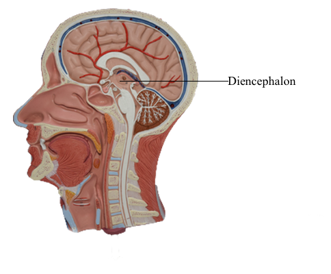
Brain : II Dicephalon
Although it is considered by some investigators to be part of
the brainstem, the diencephalon is treated as a portion of
the forebrain. The diencephalon includes the dorsal thalamus,
hypothalamus, ventral thalamus, and epithalamus, and it is situated between the telencephalon and the brainstem. In general,
the diencephalon is the main processing center for information
destined to reach the cerebral cortex from all ascending sensory pathways (except those related to olfaction). The right and left
halves of the diencephalon, for the most part, contain symmetrically distributed cell groups separated by the space of the third ventricle.
Overview
The dorsal thalamus, or thalamus as it is commonly called, is
the largest of the four principal subdivisions of the diencephalon and consists of pools of neurons that collectively project to nearly all areas of the cerebral cortex. Some of the thalamic
nuclei receive somatosensory, visual, or auditory input and
transmit this information to the appropriate area of the cerebral cortex. Other thalamic nuclei receive input from subcortical motor areas and project to those parts of the overlying
cortex that influence the successful execution of a motor act.
A few thalamic nuclei receive a more diffuse input and accordingly relate in a more diffuse way to widespread areas of the
cortex.
The hypothalamus is also composed of multiple nuclear subdivisions and is connected primarily to portions of the forebrain,
brainstem, and spinal cord. This part of the diencephalon is involved in the control of visceromotor (autonomic) functions.
In this respect, the hypothalamus regulates functions that are "automatically" adjusted (such as blood pressure and body temperature) without our being aware of the change. In contrast,
conscious sensation and some aspects of motor control are mediated by the dorsal thalamus.
The ventral thalamus and epithalamus are the smallest subdivisions of the diencephalon. The ventral thalamus includes
the subthalamic nucleus, which is linked to the basal nuclei of
the forebrain and functions in the motor sphere; lesions in the
subthalamus give rise to characteristic involuntary movement
disorders. The epithalamus is functionally related to the limbic
system.
Vasculature of the Diencephalon
The diencephalon is supplied by smaller vessels that branch from
the various arteries making up the cerebral arterial circle (circle
of Willis) and by larger arteries that originate from the proximal parts of the posterior cerebral artery.
The hypothalamus and subthalamus are supplied by central (perforating or ganglionic) branches of the circle. Anterior parts of
the hypothalamus are served by central branches (anteromedial
group) arising from the anterior communicating artery and the
A1 segment of the anterior cerebral artery and from branches of the proximal part of the posterior communicating artery. Caudal
hypothalamic regions and the ventral thalamus are supplied by
branches of the posteromedial group; these branches arise from
the posterior communicating artery and the P1 segment of the
posterior cerebral artery.
Some of the branches of the posteromedial group that arise
from the P1 segment near the basilar bifurcation are called the
thalamoperforating arteries. These vessels (of which there may
be more than one on each side) penetrate deeply to supply rostral
areas of the thalamus. If these vessels are occluded during surgery in this region, as can occur, for
example, when an aneurysm of the basilar bifurcation is clipped,
the patient can be rendered permanently comatose. Slightly
more distal branches, which usually arise from the P2 segment,
are the posterior choroidal and thalamogeniculate arteries. These
arteries also supply portions of the diencephalon. A narrow portion of the caudal and medial thalamus
bordering on the third ventricle is supplied by the medial posterior choroidal artery; the thalamogeniculate branches irrigate the
caudal thalamus, including the pulvinar and the geniculate nuclei. In addition, branches of the medial
posterior choroidal artery also serve the choroid plexus of the
third ventricle.
The anterior choroidal artery originates from the cerebral
portion of the internal carotid artery and courses caudolaterally along the trajectory of the optic tract. This
vessel serves important structures in this general area. It sends
penetrating branches into the genu of the internal capsule and
into the more inferior aspect of the posterior limb of the internal
capsule. In addition, it serves the optic tract,
inferior portions of the lenticular nucleus, the choroid plexus of
the inferior horn of the lateral ventricle, much of the amygdala,
the retrolenticular limb of the internal capsule, and large parts of
the hippocampal formation. An occlusion of this vessel, an anterior choroidal artery syndrome, results in characteristic visual
and motor deficits that reflect damage to the optic tract and the
inferior portion of the posterior limb of the internal capsule.
Although the thalamus receives a blood supply largely separate from that of the internal capsule, vascular
lesions in the thalamus may extend into the internal capsule or
vice versa. Ischemic or hemorrhagic strokes in the hemisphere may result in contralateral hemiparesis in combination with
hemianesthesia. These losses correlate with damage to corticospinal and thalamocortical fibers in the internal capsule. On
the other hand, strokes involving the larger thalamic arteries,
such as the thalamogeniculate artery, may result in total or
dissociated sensory losses. These patients may subsequently
experience persistent, intense pain (thalamic pain, DejerineRoussy syndrome).