Main Model

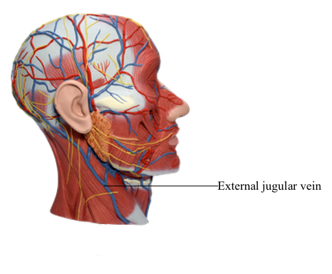
Blood Vessels : 63 External jugular vein
Lateral Cervical Region
The lateral cervical region (posterior triangle) is bounded:
• Anteriorly by the posterior border of the SCM.
• Posteriorly by the anterior border of the trapezius.
• Inferiorly by the middle third of the clavicle between the trapezius and the SCM.
• By an apex, where the SCM and trapezius meet on the superior nuchal line of the occipital bone.
• By a roof, formed by the investing layer of deep cervical fascia.
• By a floor, formed by muscles covered by the prevertebral layer of deep cervical fascia.
The lateral cervical region wraps around the lateral surface of the neck like a spiral. The region is covered by skin and subcutaneous tissue containing the platysma.
Veins in Lateral Cervical Region
The external jugular vein (EJV) begins near the angle of the mandible (just inferior to the auricle) by the union of the posterior division of the retromandibular vein with the posterior auricular vein. The EJV crosses the SCM obliquely, deep to the platysma, and enters the antero-inferior part of the lateral cervical region. It then pierces the investing layer of deep cervical fascia, which forms the roof of this region, at the posterior border of the SCM. The EJV descends to the inferior part of the lateral cervical region and terminates in the subclavian vein. It drains most of the scalp and side of the face.
The subclavian vein, the major venous channel draining the upper limb, curves through the inferior part of the lateral cervical region. It passes anterior to the anterior scalene muscle and phrenic nerve and unites at the medial border of the muscle with the IJV to form the brachiocephalic vein, posterior to the medial end of the clavicle. Just superior to the clavicle, the EJV receives the cervicodorsal, suprascapular, and anterior jugular veins.