Main Model

Brain : IV Metencephalon
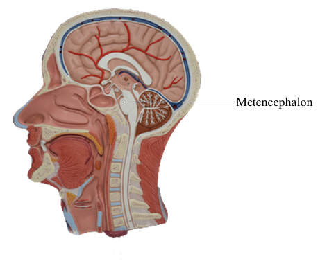
The metencephalon consists of the pons and cerebellum. The
pons is the middle segment of the brainstem, the caudal part
being the medulla and the rostral portion being the midbrain. Although comprising only about 1.3% of the brain by weight,
the pons has many important functions. The motor and sensory
nuclei and the exit points of cranial nerves V to VIII are associated with the pons. The cerebellum is not part of the brainstem
but rather is considered a suprasegmental structure because it is
located superior to the brainstem. The cerebellum is comparatively large, comprising about 10.5% of the total brain weight.
Functionally, it is part of the motor system. The blood supply to
the pons and cerebellum arises from branches of the basilar and
cerebellar arteries.
External Features
Basilar Pons
The portion of the brainstem lying between the midbrain rostrally
and the medulla caudally is the pons (pons, Latin for "bridge").
Anteriorly and laterally, the pons consists of a massive bundle of transversely oriented fibers that enter the cerebellum
as the middle cerebellar peduncle (brachium pontis). The exit of
the trigeminal nerve marks the transition from the basilar pons,
which is anterior to the trigeminal root, to the middle cerebellar
peduncle, which lies posterior to the exit of the trigeminal nerve. Rostrally, the large axonal bundles forming the crus cerebri of the midbrain extend into the basilar pons.
Caudally, some of these axons emerge to form the pyramids of
the medulla.
The cranial nerves that emerge from the pons are the trigeminal (V), abducens (VI), facial (VII), and vestibulocochlear
(VIII). The trigeminal nerve exits laterally and is composed of a large sensory root (the portio major) and a small motor root (the
portio minor). The portion of the trigeminal nerve
that traverses the subarachnoid space between the pons and
the trigeminal ganglion forms a landmark that is visible on magnetic resonance imaging at this level. The abducens,
facial, and vestibulocochlear nerves emerge in medial to lateral
sequence along the pons-medulla junction. Although
cranial nerve VII is commonly called the facial nerve, it is actually
composed of two roots, the facial nerve (SE fibers) and the intermediate nerve (VA, VE, and SA fibers). The vestibulocochlear
nerve (SA fibers) emerges posterolaterally and, with the facial
and intermediate nerves and labyrinthine artery, occupies the
internal acoustic meatus.
Rhomboid Fossa of the Pons
The rhomboid fossa forms the floor of the fourth ventricle. Its
caudal portion is located in the medulla, and its larger, more
rostral area is in the pons. The posterior surface of the pontine tegmentum, which forms the floor of the fourth ventricle, is visible only when the cerebellum is detached from the brainstem. This part of the ventricular floor is characterized
by the facial colliculus located between the median fissure and
the superior fovea of the sulcus limitans and by the vestibular
area located lateral to the sulcus limitans. The facial colliculus
is formed by the underlying abducens nucleus and internal genu
of the facial nerve, and the vestibular area marks the location
of the vestibular nuclei. The brachium pontis and the brachium conjunctivum form the lateral walls of the fourth ventricle in the
pons; the roof is formed by the anterior medullary velum, by a
small part of the cerebellum, and by a portion of the tela choroidea.
Cerebellum
The cerebellum is located posterior to the brainstem and fills
much of the posterior fossa. It is attached to the brainstem by
three pairs of cerebellar peduncles (superior, middle, and inferior). In sagittal section, the human cerebellum appears wedge
shaped, with its superior surface apposed to the tentorium cerebelli and its inferior surface curving toward the foramen magnum.
The cerebellum consists of anterior, posterior, and flocculonodular lobes; each lobe, in turn, is composed of lobules. Lobes and lobules are separated from each other
by fissures. Lobules are made up of yet smaller folds of cerebellar cortex called folia (singular, folium). Cerebellar folia, lobules,
and lobes can often be followed across the midline from one side
of the cerebellum to the other.
Each cerebellar lobe (and lobule) is also divided into rostrocaudally oriented regions of cortex commonly called the vermis
(medial), intermediate (paravermis), and hemisphere (lateral) zones. The vermal zone is approximately 1.0 cm
across at its widest point. The hemisphere is expansive in the
human cerebellum and is separated from the vermis by the intermediate zone.
Four cerebellar nuclei are located in the white matter core
of each hemisphere. From medial to lateral, they are the fastigial, globose, emboliform, and dentate nuclei. These cells receive input from branches of cerebellar afferent fibers
and from Purkinje cells located in the cerebellar cortex. In turn,
axons of cerebellar nuclear cells provide the main output signals
of the cerebellum.
Vasculature of the Pons and Cerebellum
The basilar artery and its branches serve basilar and tegmental areas of the pons. The superior cerebellar artery, through its medial and lateral branches, distributes to the superior surface of the cerebellum and most of
the cerebellar nuclei; the inferior surface of the cerebellum
is served by anterior and posterior inferior cerebellar arteries.