Main Model

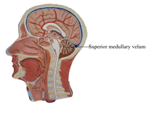
Brain : 19 Superior medullary velum
Ventricular Spaces of the Brainstem
The ventricular spaces of the brainstem are the cerebral aqueduct
in the mesencephalon and the fourth ventricle in the rhombencephalon. The cerebral aqueduct is a narrow channel, 1 to 3 mm in diameter, that connects the third ventricle
(the cavity of the diencephalon) with the fourth ventricle (the
rhombencephalic cavity). The cerebral aqueduct contains no
choroid plexus; its walls are formed by a continuous mantle of
cells collectively called the periaqueductal gray. The roof of the
midbrain is the tectum.
The fourth ventricle is the cavity of the rhombencephalon.
Its rostral portion lies between the pons and cerebellum, and its
caudal part is located in the medulla. The fourth ventricle is continuous rostrally with the cerebral aqueduct, caudally
with the central canal of the caudal medulla and cervical spinal
cord, and laterally with the subarachnoid space via the midline foramen of Magendie and the two lateral foramina of Luschka.
The foramen of Magendie is located in the caudal roof of the
ventricle and opens into the dorsal cerebellomedullary cistern (cisterna magna). The lateral recesses of the fourth
ventricle extend around the brainstem at the pons-medulla junction and end at the foramen of Luschka, which opens into the
lateral cerebellomedullary cistern.
The roof of the fourth ventricle is formed mainly by the anterior (or superior) medullary velum rostrally, by the thin
membranous tela choroidea caudally, and by a small part of the cerebellum in the middle. From rostral
to caudal, the walls of the fourth ventricle are formed by the
superior cerebellar peduncles, the middle and inferior cerebellar peduncles, and the attachment of the tela choroidea to the
medulla. The tela arises from the inferior
surface of the cerebellum and sweeps caudally to attach to the
V-shaped edges of the medullary portion of the ventricular space.
The choroid plexus of the fourth ventricle is suspended from
the inner surface of the tela, and parts of it protrude outward
through the foramina of Luschka.