Main Model

Brain : 14 Pineal gland
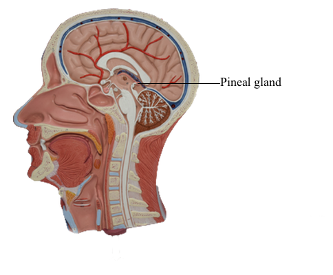
Epithalamus
The pineal gland, habenular nuclei, and stria medullaris thalami are the principal components of the epithalamus. The pineal gland consists of richly vascularized connective tissue containing glial cells and pinealocytes but no true neurons. Mammalian pinealocytes are related to the photoreceptor elements found in this gland in lower forms, such as amphibians. In humans, however, they remain only indirectly light sensitive and receive information concerning photic stimuli through a multisynaptic neural circuit.
Pinealocytes have club-like processes that are apposed to blood vessels but do not have direct synaptic contacts with central nervous system neurons. These cells synthesize melatonin from serotonin via enzymes that are sensitive to diurnal fluctuations in light. Levels of serotonin N-acetyltransferase increase during the night (in the absence of photic stimulation), and the synthesis of melatonin is enhanced. Exposure to light turns off the enzymatic activity, and melatonin production is diminished. Thus the production of melatonin by pinealocytes is rhythmic and calibrated to the 24-hour cycle of photic input to the retina. This is called a circadian rhythm.
Photic stimulation of pinealocytes occurs through an indirect route. Retinal ganglion cells project to the suprachiasmatic nucleus of the hypothalamus, which in turn influences neurons of the intermediolateral cell column in the spinal cord through descending connections. These preganglionic sympathetic neurons project to the superior cervical ganglion, which in turn innervates the pineal gland via postganglionic fibers that travel on branches of the internal carotid artery.
Pinealocytes also produce serotonin, norepinephrine, and neuroactive peptides, such as thyrotropin-releasing hormone, which are normally associated with the hypothalamus. These secretory products are released into the general circulation or the cerebrospinal fluid.
Pinealomas (tumors with large numbers of pinealocytes) are accompanied by depression of gonadal function and delayed puberty, whereas lesions that lead to the loss of pineal cells are associated with precocious puberty. This indicates that pineal secretory products exert an inhibitory influence on gonadal formation.
The habenular nuclei are located just anterior to the pineal gland and consist of a large lateral nucleus and a small medial nucleus. Both nuclei contribute axons to the habenulointerpeduncular tract (fasciculus retroflexus), which terminates in the midbrain interpeduncular nucleus. The stria medullaris thalami, which arches over the medial aspect of the dorsal thalamus near the midline, conveys input to both habenular nuclei. The habenular commissure, a small bundle of fibers riding on the upper edge of the posterior commissure, connects the habenular regions of the two sides.