Main Model

Brain : V Myelencephalon
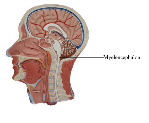
The medulla oblongata, or myelencephalon, is the most caudal
segment of the brainstem. It extends from the level of the foramen magnum to the pons. The cavity of the medulla consists of
a narrow, caudal part, which is the continuation of the central
canal of the cervical spinal cord, and a flared, rostral portion,
which is the medullary part of the fourth ventricle. The modest
size of the medulla (0.5% of total brain weight) belies its importance. All the tracts passing to or from the spinal cord traverse
the medulla, and 7 of the 12 cranial nerves (VI to XII) are associated with the medulla or the pons-medullary junction. Also, the
medullary reticular formation contains cell groups that influence
heart rate and respiration. The blood supply to the medulla arises
from branches of the vertebral arteries.
External Features
Anterior Medulla
The anterior (ventral) aspect of the medulla is characterized by
an anterior median fissure; two laterally adjacent longitudinal
ridges, the pyramids; and the olive (inferior olivary eminence). The pyramids issue from the basilar pons and extend
caudally to the motor (pyramidal) decussation, where about 90%
of their fibers cross the midline. Most of the fibers that form the pyramid arise in the motor cortex as corticospinal fibers; consequently, their crossing is called the motor decussation. Rootlets
of the hypoglossal nerve (cranial nerve XII) exit the medulla via the preolivary sulcus, a shallow groove located between the
pyramid and the olive. The abducens nerve (cranial nerve VI)
emerges at the pons-medullary junction, generally in line with
the rootlets of cranial nerve XII.
Lateral Medulla
On the lateral aspect of the medulla, a shallow trough, the postolivary sulcus or retro-olivary sulcus, is located between the restiform body and the large eminence formed by the underlying inferior olivary nucleus. Cranial nerves IX (glossopharyngeal) and X (vagus) emerge from the postolivary sulcus. Caudal rootlets of the vagus have been incorrectly called
the medullary, or bulbar, root of the accessory nerve. In actuality, the accessory nerve is made up of axons that arise from
cells in the upper levels of the cervical spinal cord (C1 to C5 or C6), ascend through the foramen magnum, coalesce to form
the accessory nerve, and then exit the skull via the jugular foramen along with the glossopharyngeal and vagus nerves. Structures served by the accessory nerve receive no innervation from
the medulla. The facial nerve (VII), along with its intermediate
root, and the vestibulocochlear nerve (VIII) emerge from the posterolateral medulla at the pons-medulla interface. The general region of the exit of the facial and vestibulocochlear nerves
is clinically regarded as the cerebellopontine angle. Indeed, a vestibular schwannoma (often incorrectly referred to as an
acoustic neuroma) is a tumor of the vestibular portion of the
eighth cranial nerve and is a lesion located at the cerebellopontine angle. On the lateral medullary surface caudal to the level
of the obex, fibers of the spinal trigeminal nucleus and tract
assume a superficial location and form the trigeminal tubercle (tuberculum cinereum). Rostral to the obex, these trigeminal fibers are located internal to a progressively
enlarging restiform body.
Posterior Medulla
At and caudal to the level of the obex, the posterior surface
of the medulla is characterized by the gracile and cuneate fasciculi and their respective tubercles. These tubercles are formed by the underlying gracile and cuneate nuclei.
Rostrolateral to the gracile and cuneate tubercles and forming a
prominent elevation on the posterolateral aspect of the medulla
is the restiform body. This structure contains a variety of afferent
cerebellar fibers and becomes progressively larger as it extends
toward the pons-medulla junction. In the caudal pons, fibers of
the restiform body join with a much smaller bundle, the juxtarestiform body, to form the inferior cerebellar peduncle.
Vasculature
In general, the blood supply to the entire medulla and to the choroid plexus of the fourth ventricle arises from branches of the vertebral arteries. The exceptions are the portion of
the choroid plexus that extends out of the foramen of Luschka and
the adjacent cochlear nuclei; these are served by branches of the
anterior inferior cerebellar artery, a branch of the basilar artery. In
general, the medial medulla is served by the anterior spinal artery,
the anterolateral medulla by small branches from the vertebral
artery, and the posterolateral medulla rostral to the obex by the
posterior inferior cerebellar artery (PICA). Caudal to the obex,
the posterior medulla is served by the posterior spinal artery.
The vascular territory of the anterior spinal artery encompasses
the medial lemniscus, hypoglossal root, and corticospinal fibers in
the pyramid. Consequently, lesions of this vascular region result
in somatosensory and motor deficits reflecting damage to these
structures. In similar manner, lesions in the territory of the PICA will damage the anterolateral system and spinal trigeminal tract
and nucleus and give rise to deficits reflecting these structures.