Main Model

Larynx : XI Esophagus
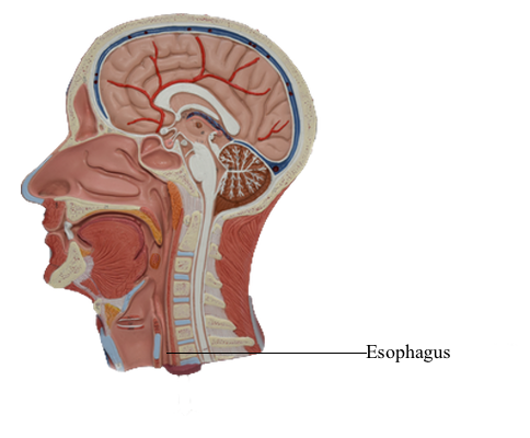
The esophagus is a muscular tube that connects the pharynx to the stomach. It begins in the neck where it is continuous with the laryngopharynx at the pharyngo-esophageal junction. The esophagus consists of striated (voluntary) muscle in its upper third, smooth (involuntary) muscle in its lower third, and a mixture of striated and smooth muscle in between.
Its first part, the cervical esophagus, is part of the voluntary upper third. It begins immediately posterior to, and at the level of, the inferior border of the cricoid cartilage in the median plane. This is the level of the C6 vertebra.
Externally, the pharyngo-esophageal junction appears as a constriction produced by the cricopharyngeal part of the inferior pharyngeal constrictor muscle (the superior esophageal sphincter) and is the narrowest part of the esophagus. The cervical esophagus inclines slightly to the left as it descends and enters the superior mediastinum via the superior thoracic aperture, where it becomes the thoracic esophagus.
When the esophagus is empty, it is a slit-like lumen. When a food bolus descends in it, the lumen expands, eliciting reflex peristalsis in the inferior two thirds of the esophagus. The cervical esophagus lies between the trachea and the cervical vertebral column. It is attached to the trachea by loose connective tissue. The recurrent laryngeal nerves lie in or near the tracheo-esophageal grooves between the trachea and esophagus. On the right of the esophagus is the right lobe of the thyroid gland and the right carotid sheath and its contents.
The esophagus is in contact with the cervical pleura at the root of the neck. On the left is the left lobe of the thyroid gland and the left carotid sheath. The thoracic duct adheres to the left side of the esophagus and lies between the pleura and the esophagus.