Main Model

Brain : 8 Interthalamic adhesion
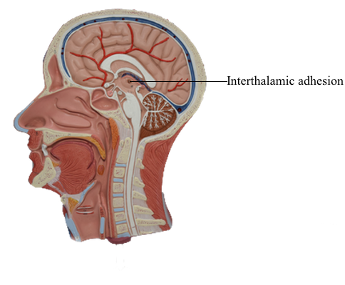
Thalamus
The gray matter of the diencephalon develops from a continuation of the brainstem alar plates; there is no homologue of the
basal plate in the diencephalon. This alar plate is recognizable at 4 weeks of gestation; by 6 weeks of gestation, it has differentiated into three main areas of the diencephalon: the epithalamus,
thalamus (dorsal thalamus), and hypothalamus.
These structures are visible as swellings in the wall of the third
ventricle, where they are separated from each other by the epithalamic and hypothalamic sulci. As development progresses,
the epithalamic area remains small, whereas the hypothalamus
and especially the thalamus enlarge. In about 80% of individuals,
the two thalami fuse across the third ventricle to form the interthalamic adhesion (massa intermedia).
The basic concepts of the development of the thalamus are the
same as for other CNS regions. Radial glia extend from the third
ventricle to the pial surface, and developing neurons migrate along this guide. The development of the thalamus occurs in an "outside-first" sequence. That is, the first neurons to undergo their final cell division migrate to the outermost portion of the thalamus, where they mature. This means that the
most lateral of the thalamic nuclei, such as the geniculate nuclei
and the lateral and ventral nuclei, are generated first. The most
medial thalamic nuclei, such as the dorsomedial nucleus, are the
last to develop.
An important process in the development of the thalamic relay
nuclei is the establishment of orderly maps of the sensory world.
For example, a retinotopic (vision) map is formed in the lateral
geniculate nucleus. As retinal ganglion cells send axons to the
lateral geniculate nucleus, the arrangement of axonal contacts on
cells in this visual relay center must accurately reflect the positions of ganglion cells in the retina. In this way, the map of visual
space on the retina is maintained in the lateral geniculate nucleus
and ultimately in the visual cortex. Similar maps are formed in
the medial geniculate nucleus (tonotopic mapping) and ventral
posterolateral nucleus (somatotopic mapping).