Main Model

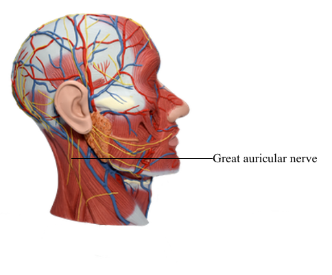
Blood Vessels : g Great auricular nerve
Nerves of Face and Scalp
Cutaneous (sensory) innervation of the face and anterosuperior part of the scalp is provided primarily by the trigeminal nerve (CN V), whereas motor innervation to the facial muscles is provided by the facial nerve (CN VII).
Cutaneous Nerves of Face and Scalp
The trigeminal nerve (CN V) originates from the lateral surface of the pons of the midbrain by two roots: motor and sensory. These roots are comparable to the motor (anterior) and sensory (posterior) roots of spinal nerves. The sensory root of CN V consists of the central processes of pseudounipolar neurons located in a sensory ganglion (trigeminal ganglion) at the distal end of the root, which is bypassed by the multipolar neuronal axons making up the motor root. CN V is the sensory nerve for the face and the motor nerve for the muscles of mastication and several small muscles.
The peripheral processes of the neurons of the trigeminal ganglion constitute three divisions of the nerve: the ophthalmic nerve (CN V1), the maxillary nerve (CN V2), and the sensory component of the mandibular nerve (CN V3). These nerves are named according to their main areas of termination: the eye, maxilla, and mandible, respectively. The first two divisions (ophthalmic and maxillary nerves) are wholly sensory. The mandibular nerve is largely sensory, but it also receives the motor fibers (axons) from the motor root of CN V that mainly supply the muscles of mastication.
The cutaneous nerves of the neck overlap those of the face. Cutaneous branches of cervical nerves from the cervical plexus extend over the posterior aspect of the neck and scalp. The great auricular nerve in particular innervates the inferior aspect of the auricle (external ear) and much of the parotid region of the face (the area overlying the angle of the jaw).
Ophthalmic Nerve
The ophthalmic nerve (CN V1), the superior division of the trigeminal nerve, is the smallest of the three divisions of CN V. It arises from the trigeminal ganglion as a wholly sensory nerve and supplies the area of skin derived from the embryonic frontonasal prominence. As CN V1 enters the orbit through the superior orbital fissure, it trifurcates into the frontal, nasociliary, and lacrimal nerves. Except for the external nasal nerve, the cutaneous branches of CN V1 reach the skin of the face via the orbital opening.
The frontal nerve, the largest branch produced by the trifurcation of CN V1, runs along the roof of the orbit toward the orbital opening, bifurcating approximately midway into the cutaneous supra-orbital and supratrochlear nerves, distributed to the forehead and scalp.
The nasociliary nerve, the intermediate branch of the CN V1 trifurcation, supplies branches to the eyeball and divides within the orbit into the posterior ethmoidal, anterior ethmoidal, and infratrochlear nerves. The posterior and anterior ethmoidal nerves leave the orbit, the latter running a circuitous course passing through the cranial and nasal cavities. Its terminal branch, the external nasal nerve, is a cutaneous nerve supplying the external nose. The infratrochlear nerve is a terminal branch of the nasociliary nerve and its main cutaneous branch.
The lacrimal nerve, the smallest branch from the trifurcation of CN V1, is primarily a cutaneous branch, but it also conveys some secretomotor fibers, sent via a communicating branch, from a ganglion associated with the maxillary nerve for innervation of the lacrimal gland.
Maxillary Nerve
The maxillary nerve (CN V2), the intermediate division of the trigeminal nerve, also arises as a wholly sensory nerve. CN V2 passes anteriorly from the trigeminal ganglion and leaves the cranium through the foramen rotundum in the base of the greater wing of the sphenoid. The maxillary nerve enters the pterygopalatine fossa, where it gives off branches to the pterygopalatine ganglion and continues anteriorly, entering the orbit through the inferior orbital fissure. It gives off the zygomatic nerve and passes anteriorly into the infraorbital groove and foramen as the infra-orbital nerve.
The zygomatic nerve runs to the lateral wall of the orbit, giving rise to two of the three cutaneous branches of CN V2, the zygomaticofacial and zygomaticotemporal nerves. The latter nerve sends a communicating branch conveying secretomotor fibers to the lacrimal nerve. En route to the face, the infra-orbital nerve gives off palatine branches, branches to the mucosa of the maxillary sinus, and branches to the posterior teeth. It reaches the skin of the face by traversing the infra-orbital foramen on the infra-orbital surface of the maxilla. The three cutaneous branches of the maxillary nerve supply the area of skin derived from the embryonic maxillary prominences.
Mandibular Nerve
The mandibular nerve (CN V3) is the inferior and largest division of the trigeminal nerve. It is formed by the union of sensory fibers from the sensory ganglion and the motor root of CN V in the foramen ovale in the greater wing of the sphenoid, through which CN V3 emerges from the cranium. CN V3 has three sensory branches that supply the area of skin derived from the embryonic mandibular prominence. It also supplies motor fibers to the muscles of mastication. CN V3 is the only division of CN V that carries motor fibers. The major cutaneous branches of CN V3 are the auriculotemporal, buccal, and mental nerves. En route to the skin, the auriculotemporal nerve passes deep to the parotid gland, conveying secretomotor fibers to it from a ganglion associated with this division of CN V.
Nerves of Scalp
Innervation of the scalp anterior to the auricles of the external ears is through branches of all three divisions of CN V, the trigeminal nerve. Posterior to the auricles, the nerve supply is from spinal cutaneous nerves (C2 and C3).
Motor Nerves of Face
The motor nerves of the face are the facial nerve to the muscles of facial expression and the motor root of the trigeminal nerve/mandibular nerve to the muscles of mastication (masseter, temporal, medial, and lateral pterygoids). These nerves also supply some more deeply placed muscles.
Facial Nerve
CN VII, the facial nerve, has a motor root and a sensory/ parasympathetic root (the latter being the intermediate nerve). The motor root of CN VII supplies the muscles of facial expression, including the superficial muscle of the neck (platysma), auricular muscles, scalp muscles, and certain other muscles derived from mesoderm in the embryonic second pharyngeal arch. Following a circuitous route through the temporal bone, CN VII emerges from the cranium through the stylomastoid foramen located between the mastoid and styloid processes. It immediately gives off the posterior auricular nerve, which passes posterosuperior to the auricle of the ear to supply the auricularis posterior and occipital belly of the occipitofrontalis muscle.
The main trunk of CN VII runs anteriorly and is engulfed by the parotid gland, in which it forms the parotid plexus. This plexus gives rise to the five terminal branches of the facial nerve: temporal, zygomatic, buccal, marginal mandibular, and cervical. The names of the branches refer to the regions they supply.
The temporal branch of CN VII emerges from the superior border of the parotid gland and crosses the zygomatic arch to supply the auricularis superior and auricularis anterior; the frontal belly of the occipitofrontalis; and, most important, the superior part of the orbicularis oculi.
The zygomatic branch of CN VII passes via two or three branches superior and mainly inferior to the eye to supply the inferior part of the orbicularis oculi and other facial muscles inferior to the orbit.
The buccal branch of CN VII passes external to the buccinator to supply this muscle and the muscles of the upper lip (upper parts of orbicularis oris and inferior fibers of levator labii superioris).
The marginal mandibular branch of CN VII supplies the risorius and muscles of the lower lip and chin. It emerges from the inferior border of the parotid gland and crosses the inferior border of the mandible deep to the platysma to reach the face. In approximately 20% of people, this branch passes inferior to the angle of the mandible.
The cervical branch of CN VII passes inferiorly from the inferior border of the parotid gland and runs posterior to the mandible to supply the platysma.