Main Model

Brain : I Telencephalon
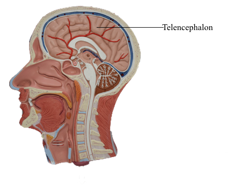
The telencephalon is the largest part of the human brain, constituting about 85% of total brain weight, and is that portion in
which all modalities are represented. Various sensory inputs (such
as vision and hearing) are localized in some areas, whereas motor
functions are represented in other regions and are modulated
by subcortical nuclei. The telencephalon contains circuits that
interrelate regions that have specific functions, such as motor
or visual, with other regions called association areas. Seeing a
familiar image may precipitate a cascade of neural events having
olfactory, emotional, sensory, and motor components. Damage
to association areas results in complex neurologic deficits. The
patient may not be blind or paralyzed but may be unable to recognize sensory input (agnosia), express ideas or thoughts (aphasia), or perform complex goal-directed movements (apraxia).
Overview
The telencephalon consists of large hemispheres separated from
each other by a deep longitudinal cerebral fissure. Each hemisphere has an outer surface, the cerebral cortex, which is composed of layers of cells. The cortex is thrown into elevations
called gyri (singular, gyrus) that are separated by grooves called
sulci (singular, sulcus). Internal to the cortex are large amounts
of subcortical white matter along with aggregates of gray matter
that form the basal nuclei and the amygdala. Although not parts
of either the telencephalon or the basal nuclei, the subthalamic
nucleus (of the diencephalon) and the substantia nigra (of the
mesencephalon) have important connections that functionally
link them with the basal nuclei.
Information passing into or out of the cerebral cortex must
traverse the subcortical white matter. The myelinated fibers
forming the white matter are organized into (1) association bundles that connect adjacent or distant gyri in one hemisphere;
(2) commissural bundles that connect the hemispheres, the largest of these being the corpus callosum; and (3) the internal capsule. The internal capsule contains axons projecting to numerous
downstream nuclei (corticofugal fibers) and axons conveying
information to the cerebral cortex (corticopetal fibers). The
terms corticofugal and corticopetal are umbrella terms that
include all efferent and all afferent fibers, respectively, of the
cerebral cortex.
The hippocampal complex and the amygdala are located in
the walls of the temporal horn of the lateral ventricle. The axons
of cells in these structures coalesce to form the fornix, stria terminalis, and amygdalofugal pathway.