Main Model

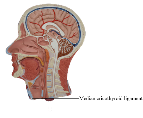
Larynx : V Median cricothyroid ligament
The posterior border of each lamina projects superiorly as the superior
horn and inferiorly as the inferior horn. The superior border and
superior horns attach to the hyoid by the thyrohyoid membrane. The thick
median part of this membrane is the median thyrohyoid ligament; its lateral parts are the lateral thyrohyoid ligaments.
The inferior horns articulate with the lateral surfaces of the cricoid cartilage at the cricothyroid joints. The main movements at these joints are rotation and gliding of the thyroid cartilage, which result in changes in the length of the vocal folds. The cricoid cartilage is shaped like a signet ring with its band facing anteriorly. This ring-like opening of the cartilage fits an average finger. The posterior (signet) part of the cricoid is the lamina, and the anterior (band) part is the arch. Although much smaller than the thyroid cartilage, the cricoid cartilage is thicker and stronger and is the only complete ring of cartilage to encircle any part of the airway. It attaches to the inferior margin of the thyroid cartilage by the median cricothyroid ligament and to the first tracheal ring by the cricotracheal ligament. Where the larynx is closest to the skin and most accessible, the median cricothyroid ligament may be felt as a soft spot during palpation inferior to the thyroid cartilage.