Main Model

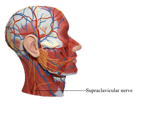
Blood Vessels : l Supraclaviclar nerve
Lateral Cervical Region
The lateral cervical region (posterior triangle) is bounded:
• Anteriorly by the posterior border of the SCM.
• Posteriorly by the anterior border of the trapezius.
• Inferiorly by the middle third of the clavicle between the trapezius and the SCM.
• By an apex, where the SCM and trapezius meet on the superior nuchal line of the occipital bone.
• By a roof, formed by the investing layer of deep cervical fascia.
• By a floor, formed by muscles covered by the prevertebral layer of deep cervical fascia.
The lateral cervical region wraps around the lateral surface of the neck like a spiral. The region is covered by skin and subcutaneous tissue containing the platysma.
Nerves of Lateral Cervical Region
The spinal accessory nerve (CN XI) passes deep to the SCM, supplying it before entering the lateral cervical region at or inferior to the junction of the superior and middle thirds of the posterior border of the SCM. The nerve passes postero-inferiorly, within or deep to the investing layer of deep cervical fascia, running on the levator scapulae from which it is separated by the prevertebral layer of fascia. CN XI then disappears deep to the anterior border of the trapezius at the junction of its superior two thirds with its inferior one third.
The roots of the brachial plexus (anterior rami of C5-C8 and T1) appear between the anterior and the middle scalene muscles. The five rami unite to form the three trunks of the brachial plexus, which descend inferolaterally through the lateral cervical region. The plexus then passes between the 1st rib, clavicle, and superior border of the scapula (the cervico-axillary canal) to enter the axilla, providing innervation for most of the upper limb.
The suprascapular nerve, which arises from the superior trunk of the brachial plexus (not cervical plexus), runs laterally across the lateral cervical region to supply the supraspinatus and infraspinatus muscles on the posterior aspect of the scapula. It also sends articular branches to the glenohumeral joint.
The anterior rami of C1-C4 make up the roots of the cervical plexus. The cervical plexus consists of an irregular series of (primary) nerve loops and the branches that arise from the loops. Each participating ramus, except the first, divides into ascending and descending branches that unite with the branches of the adjacent spinal nerve to form the loops. The cervical plexus lies anteromedial to the levator scapulae and middle scalene muscles and deep to the SCM. The superficial branches of the plexus that initially pass posteriorly are cutaneous (sensory) branches. The deep branches passing anteromedially are motor branches, including the roots of the phrenic nerve (to the diaphragm) and the ansa cervicalis.
The superior root of the ansa cervicalis, conveying fibers from spinal nerves C1 and C2, briefly joins and then descends from the hypoglossal nerve (CN XII) as it traverses the lateral cervical region. The inferior root of the ansa cervicalis arises from a loop between spinal nerves C2 and C3. The superior and inferior roots of the ansa cervicalis unite, forming a secondary loop, the ansa cervicalis, consisting of fibers from the C1-C3 spinal nerves, which branch from the secondary loop to supply the infrahyoid muscles, including the omohyoid, sternothyroid, and sternohyoid. The fourth infrahyoid muscle, the thyrohyoid, receives C1 fibers, which descend independently from the hypoglossal nerve, distal to the superior root of the ansa cervicalis (nerve to thyrohyoid).
Cutaneous branches of the cervical plexus emerge around the middle of the posterior border of the SCM, often called the nerve point of the neck, and supply the skin of the neck, superolateral thoracic wall, and scalp between the auricle and the external occipital protuberance. Close to their origin, the roots of the cervical plexus receive gray rami communicantes, most of which descend from the large superior cervical ganglion in the superior part of the neck.
Branches of cervical plexus arising from the nerve loop between the anterior rami of C2 and C3 are the:
• Lesser occipital nerve (C2): supplies the skin of the neck and scalp posterosuperior to the auricle.
• Great auricular nerve (C2 and C3): ascends vertically across the oblique SCM to the inferior pole of the parotid gland, where it divides to supply the skin over - and the sheath surrounding - the gland, the mastoid process, and both surfaces of the auricle and an area of skin extending from the angle of the mandible to the mastoid process.
• Transverse cervical nerve (C2 and C3): supplies the skin covering the anterior cervical region. It curves around the middle of the posterior border of the SCM inferior to the great auricular nerve and passes anteriorly and horizontally across it deep to the EJV and platysma, dividing into superior and inferior branches.
The branches of the cervical plexus arising from the nerve loop formed between the anterior rami of C3-C4 are the:
• Supraclavicular nerves (C3 and C4): emerge as a common trunk under cover of the SCM, sending small branches to the skin of the neck that cross the clavicle and supply the skin over the shoulder.
In addition to the ansa cervicalis and phrenic nerves arising from the loops of the plexus, deep motor branches of the cervical plexus include branches arising from the roots that supply the rhomboids (dorsal scapular nerve; C4 and C5), serratus anterior (long thoracic nerve; C5-C7), and nearby prevertebral muscles.
The phrenic nerves originate chiefly from the C4 nerve but receive contributions from the C3 and C5 nerves. The phrenic nerves contain motor, sensory, and sympathetic nerve fibers. These nerves provide the sole motor supply to the diaphragm as well as sensation to its central part. In the thorax, each phrenic nerve supplies the mediastinal pleura and pericardium. Receiving variable communicating fibers in the neck from the cervical sympathetic ganglia or their branches, each phrenic nerve forms at the superior part of the lateral border of the anterior scalene muscle at the level of the superior border of the thyroid cartilage. The phrenic nerve descends obliquely with the IJV across the anterior scalene, deep to the prevertebral layer of deep cervical fascia and the transverse cervical and suprascapular arteries.
On the left, the phrenic nerve crosses anterior to the first part of the subclavian artery; on the right, it lies on the anterior scalene muscle and crosses anterior to the second part of the subclavian artery. On both sides, the phrenic nerve runs posterior to the subclavian vein and anterior to the internal thoracic artery as it enters the thorax.
The contribution of the C5 nerve to the phrenic nerve may be derived from an accessory phrenic nerve. Frequently, it is a branch of the nerve to the subclavius. If present, the accessory phrenic nerve lies lateral to the main nerve and descends posterior and sometimes anterior to the subclavian vein. The accessory phrenic nerve joins the phrenic nerve either in the root of the neck or in the thorax.