Main Model

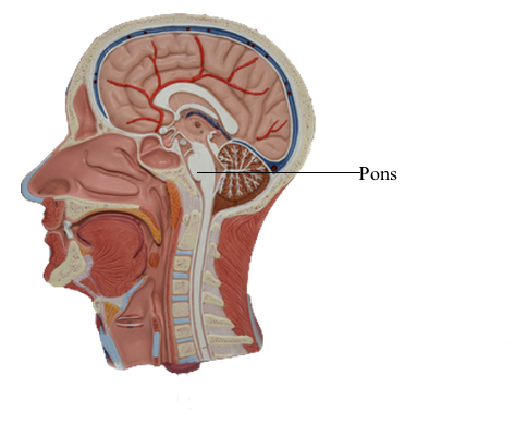
Brain : 20 Pons
The term brainstem (sometimes written brain stem) can mean
either the portion of the brain that consists of the medulla oblongata, pons, and midbrain or the portion that consists of these structures plus the diencephalon. For our purposes, therefore, the brainstem consists
of the rhombencephalon (excluding the cerebellum) and the
mesencephalon. These regions of the brainstem share a basic
organization.
Pons
The pons is the part of the brainstem between the midbrain rostrally and the medulla oblongata caudally; it lies in the anterior part of the posterior cranial fossa. CN V is associated with the pons. The pons (the anterior part of the metencephalon) extends from
the pons-medulla junction to an imaginary line drawn from the
exit of the trochlear nerve posteriorly to the rostral edge of the
basilar pons anteriorly. What we commonly call
the pons is actually composed of two portions, the pontine tegmentum (located internally) and the basilar pons. The basilar pons is bulbous and quite characteristic of the anterior
aspect of the pons. The pontine tegmentum contains portions of
the trigeminal nuclei and the vestibular nuclei and, just rostral
to the pons-medulla junction, the facial motor nucleus, superior salivatory nucleus, and abducens nucleus. The trigeminal
nerve (V, mixed) emerges from the lateral aspect of the pons, and
abducens (VI), facial (VII), and vestibulocochlear (VIII) nerves
exit at the pons-medulla junction.
The cerebellum, although part of the metencephalon, is not
part of the brainstem. It is joined to the brainstem by three large,
paired bundles of fibers called the cerebellar peduncles. These are
the inferior cerebellar peduncle, the middle cerebellar peduncle (or brachium pontis), and the superior cerebellar peduncle
(or brachium conjunctivum), connecting the cerebellum to the medulla oblongata, basilar pons, and midbrain, respectively.
Tegmental and Basilar Areas
The central core of the midbrain and the pons is called the tegmentum, and their anterior (ventral) parts are the basilar areas.
These regions are continuous with each other and with comparable areas of the medulla. The tegmentum of the pons and midbrain and the contiguous central portion
of the medulla contain ascending and descending tracts, many
relay nuclei, and the nuclei of cranial nerves III to XII.
The basilar part of each brainstem division is anterior to the
tegmentum (of the midbrain and pons) and to the central portion of the medulla. Consequently, these basilar structures also form a rostrocaudal continuum. Basilar structures of
the brainstem include the descending fibers of the crus cerebri
(midbrain), basilar pons, and pyramid (medulla) and specific populations of neurons in the midbrain and pons that originate
from the alar plate of the embryonic brain.
External Features
Basilar Pons
The portion of the brainstem lying between the midbrain rostrally
and the medulla caudally is the pons (pons, Latin for "bridge").
Anteriorly and laterally, the pons consists of a massive bundle of transversely oriented fibers that enter the cerebellum
as the middle cerebellar peduncle (brachium pontis). The exit of
the trigeminal nerve marks the transition from the basilar pons,
which is anterior to the trigeminal root, to the middle cerebellar
peduncle, which lies posterior to the exit of the trigeminal nerve. Rostrally, the large axonal bundles forming the crus cerebri of the midbrain extend into the basilar pons.
Caudally, some of these axons emerge to form the pyramids of
the medulla.
The cranial nerves that emerge from the pons are the trigeminal (V), abducens (VI), facial (VII), and vestibulocochlear
(VIII). The trigeminal nerve exits laterally and is composed of a large sensory root (the portio major) and a small motor root (the
portio minor). The portion of the trigeminal nerve
that traverses the subarachnoid space between the pons and
the trigeminal ganglion forms a landmark that is visible on magnetic resonance imaging at this level. The abducens,
facial, and vestibulocochlear nerves emerge in medial to lateral
sequence along the pons-medulla junction. Although
cranial nerve VII is commonly called the facial nerve, it is actually
composed of two roots, the facial nerve (SE fibers) and the intermediate nerve (VA, VE, and SA fibers). The vestibulocochlear
nerve (SA fibers) emerges posterolaterally and, with the facial
and intermediate nerves and labyrinthine artery, occupies the
internal acoustic meatus.
Rhomboid Fossa of the Pons
The rhomboid fossa forms the floor of the fourth ventricle. Its
caudal portion is located in the medulla, and its larger, more
rostral area is in the pons. The posterior surface of the pontine tegmentum, which forms the floor of the fourth ventricle, is visible only when the cerebellum is detached from the brainstem. This part of the ventricular floor is characterized
by the facial colliculus located between the median fissure and
the superior fovea of the sulcus limitans and by the vestibular
area located lateral to the sulcus limitans. The facial colliculus
is formed by the underlying abducens nucleus and internal genu
of the facial nerve, and the vestibular area marks the location
of the vestibular nuclei. The brachium pontis and the brachium conjunctivum form the lateral walls of the fourth ventricle in the
pons; the roof is formed by the anterior medullary velum, by a
small part of the cerebellum, and by a portion of the tela choroidea.
Vasculature (Internal) of the Pons
Internal areas of the tegmental and basilar pons are served
by branches of the basilar artery. Paramedian
branches distribute to medial areas of the basilar pons, including
corticospinal fibers and the exiting fibers of the abducens nerve.
The lateral part of the basilar pons is served by short circumferential branches, and the entire tegmental area plus a wedge
of the middle cerebellar peduncle receives blood via the long
circumferential branches. At caudal levels (levels of the facial
colliculus), the long circumferential supply is supplemented by branches of the anterior inferior cerebellar artery. Rostrally,
beginning at about the level of the principal sensory and motor
trigeminal nuclei, the blood supply to the pontine tegmentum
is supplemented by branches of the superior cerebellar artery.