Main Model

Larynx : IV Median thyrohyoid ligament
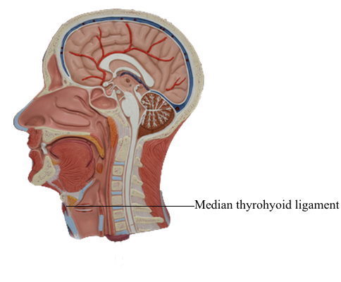
The thyroid cartilage is the largest of the cartilages; its superior border lies opposite the C4 vertebra. The inferior two thirds of its two plate-like laminae fuse anteriorly in the median plane to form the laryngeal prominence. This projection (“Adam’s apple”) is well marked in men but seldom visible in women. Superior to this prominence, the laminae diverge to form a V-shaped superior thyroid notch. The less distinct inferior thyroid notch is a shallow indentation in the middle of the inferior border of the cartilage.
The posterior border of each lamina projects superiorly as the superior horn and inferiorly as the inferior horn. The superior border and superior horns attach to the hyoid by the thyrohyoid membrane. The thick median part of this membrane is the median thyrohyoid ligament; its lateral parts are the lateral thyrohyoid ligaments.