Main Model

Brain : 15 Optic chiasm
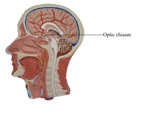
Optic Nerve, Chiasm, and Tract
Axons of retinal ganglion cells conveying input from all areas of
the retina converge at the optic disc, where they penetrate the
choroid and sclera to form the optic nerve. Within the nerve fiber
layer of the retina, ganglion cell axons are unmyelinated. However, as they pass through the sclera, they become ensheathed
with myelin formed by oligodendrocytes. Because there are no
photoreceptor cells in the optic disc (only ganglion cell axons),
light striking this area is not perceived. Consequently, this part
of the retina is commonly called the blind spot.
Visual acuity is greatest at the fovea, but the peripheral retina has
little form vision; fine details cannot be perceived in the peripheral retina because the "pixel density" (density of rod photoreceptors) is much lower.
The optic nerve extends from the caudal aspect of the eye to
the optic chiasm. This nerve is enclosed in a sleeve of
dura and arachnoid mater that is continuous with the same layers around the brain. Thus the subarachnoid space extends along the
optic nerve, which is bathed in cerebrospinal fluid. For this reason, increases in intracranial pressure may be transmitted along
the optic nerves and can cause blockage of axoplasmic flow at the
optic nerve head. This axoplasmic stasis results in swelling of the
optic nerve head (papilledema). The damage to
the optic nerve may result in partial or complete loss of vision in
that eye.
Terminal branches of the central retinal artery, a branch of the
ophthalmic artery, issue from the optic disc and radiate over the
retina. Examination of these vessels through an ophthalmoscope
can help assess the health of the eye and the central nervous
system. Changes in the configuration of the
retinal vessels or in the size or shape of the optic disc may indicate diseases of the retina, the vascular system, or the central
nervous system.
Just rostral to the pituitary stalk, the optic nerves come
together to form the optic chiasm, from which the optic tracts
diverge as they pass caudally. In the chiasm, the fibers from the nasal half of each retina (corresponding to the temporal hemifields) cross to enter the contralateral optic tract, whereas the
fibers from the temporal half of each retina (corresponding to
the nasal hemifields) remain on the same side and enter the ipsilateral optic tract. In this way, each half of the brain receives the
fibers corresponding to the contralateral half of the visual world.
Although many clinical events can affect the optic chiasm,
this structure is especially susceptible to tumors of the pituitary
gland. Enlarging pituitary tumors that damage the crossing fibers
in the midline of the chiasm will interrupt visual input from the
temporal halves of both visual fields, resulting in a bitemporal
hemianopia. A lesion that damages
the lateral part of the chiasm may interrupt only fibers conveying
information from the nasal visual field on the same side, although
in practice this situation is rare. This deficit is called an ipsilateral (right or left) nasal hemianopia.
Extending caudolaterally from the chiasm, the axons of retinal ganglion cells continue as a compact bundle, the optic tract.
This structure courses over the surface of the crus cerebri at its
junction with the hemisphere and ends in the lateral geniculate
nucleus of the diencephalon. Because the optic
tract contains fibers conveying visual input from the ipsilateral
nasal hemifield and the contralateral temporal hemifield, lesions
of the optic tract result in a contralateral (either right or left)
homonymous hemianopia.
The optic chiasm receives blood from the small anteromedial
branches of the anterior communicating artery and A1 segment
of the anterior cerebral artery. The optic nerve receives its blood
supply from small branches of the ophthalmic artery traveling
parallel to the nerve. The optic nerve head and retina are supplied by the central artery of the retina. The
optic tract receives its main blood supply from the anterior choroidal artery, whereas the lateral
geniculate nucleus is in the domain of the thalamogeniculate
artery, a branch of the posterior cerebral artery.