Main Model

Brain : 11 Interventricular foramen
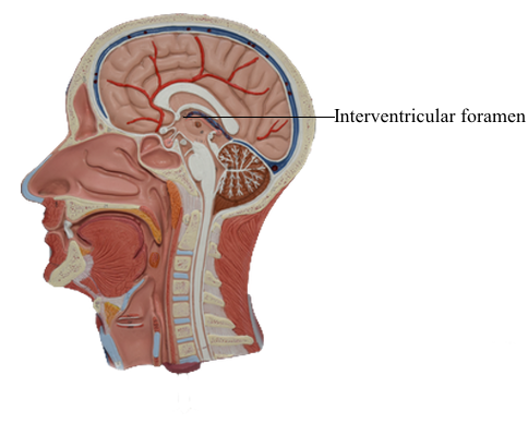
The ventricular system of the brain consists of two lateral ventricles and the midline 3rd and 4th ventricles connected by the cerebral aqueduct. CSF, largely secreted by the choroid plexuses of the ventricles, fills these brain cavities and the subarachnoid space of the brain and spinal cord.
The lateral ventricles, the 1st and 2nd ventricles, are the largest cavities of the ventricular system and occupy large areas of the cerebral hemispheres. Each lateral ventricle opens through an interventricular foramen into the 3rd ventricle. The 3rd ventricle, a slit-like cavity between the right and the left halves of the diencephalon, is continuous posteroinferiorly with the cerebral aqueduct, a narrow channel in the midbrain connecting the 3rd and 4th ventricles.
The pyramid-shaped 4th ventricle in the posterior part of the pons and medulla extends inferoposteriorly. Inferiorly, it tapers to a narrow channel that continues into the cervical region of the spinal cord as the central canal. CSF drains into the subarachnoid space from the 4th ventricle through a single median aperture and paired lateral apertures. These apertures are the only means by which CSF enters the subarachnoid space. If they are blocked, CSF accumulates and the ventricles distend, producing compression of the substance of the cerebral hemispheres.