Main Model

Larynx : II Thyroid cartilage
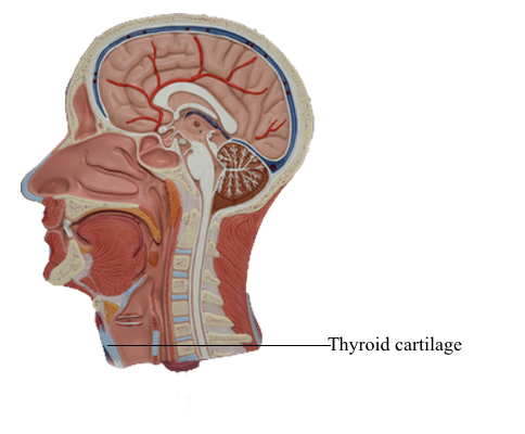
The thyroid cartilage is the largest of the cartilages; its superior border lies opposite the C4 vertebra. The inferior two thirds of its two plate-like laminae fuse anteriorly in the median plane to form the laryngeal prominence. This projection (“Adam’s apple”) is well marked in men but seldom visible in women. Superior to this prominence, the laminae diverge to form a V-shaped superior thyroid notch. The less distinct inferior thyroid notch is a shallow indentation in the middle of the inferior border of the cartilage.
The U-shaped hyoid bone lies in the anterior part of the neck in the deep angle between the mandible and the thyroid cartilage at the level of the C3 vertebra. Swallow, and the hyoid will move under your fingers when they are placed at the angle between the chin and anterior neck. The greater horn of one side of the hyoid is palpable only when the greater horn on the opposite side is steadied.
The laryngeal prominence is produced by the meeting of the laminae of the thyroid cartilage at an acute angle in the anterior midline. This thyroid angle, most acute in postpubertal males, forms the laryngeal prominence (“Adam’s apple”), which is palpable and frequently visible. During palpation of the prominence, it can be felt to recede on swallowing. The vocal folds are at the level of the middle of the laryngeal prominence.