Main Model

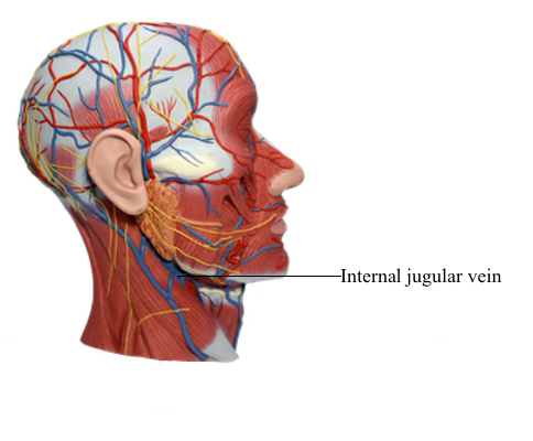
Blood Vessels : 64 Internal jugular vein
Anterior Cervical Region
The anterior cervical region (anterior triangle) has the following:
• An anterior boundary formed by the median line of the neck.
• A posterior boundary formed by the anterior border of the SCM.
• A superior boundary formed by the inferior border of the mandible.
• An apex located at the jugular notch in the manubrium.
• A roof formed by subcutaneous tissue containing the platysma.
• A floor formed by the pharynx, larynx, and thyroid gland.
For more precise localization of structures, the anterior cervical region is subdivided into four smaller triangles by the digastric and omohyoid muscles: the unpaired submental triangle and three small paired triangles - submandibular, carotid, and muscular.
The submental triangle, inferior to the chin, is a suprahyoid area bounded inferiorly by the body of the hyoid and laterally by the right and left anterior bellies of the digastric muscles. The floor of the submental triangle is formed by the two mylohyoid muscles, which meet in a median fibrous raphe. The apex of the submental triangle is at the mandibular symphysis, the site of union of the halves of the mandible during infancy. The base of the submental triangle is formed by the hyoid. This triangle contains several small submental lymph nodes and small veins that unite to form the anterior jugular vein.
The submandibular triangle is a glandular area between the inferior border of the mandible and the anterior and posterior bellies of the digastric muscle. The floor of the submandibular triangle is formed by the mylohyoid and hyoglossus muscles, and the middle pharyngeal constrictor. The submandibular gland nearly fills this triangle.
Submandibular lymph nodes lie on each side of the submandibular gland and along the inferior border of the mandible. The hypoglossal nerve (CN XII) provides motor innervation to the intrinsic and extrinsic muscles of the tongue. It passes into the submandibular triangle, as does the nerve to the mylohyoid (a branch of CN V3, which also supplies the anterior belly of the digastric), parts of the facial artery and vein, and the submental artery (a branch of the facial artery).
The carotid triangle is a vascular area bounded by the superior belly of the omohyoid, the posterior belly of the digastric, and the anterior border of the SCM. This triangle is important because the common carotid artery ascends into it. Its pulse can be auscultated or palpated by compressing it lightly against the transverse processes of the cervical vertebrae. At the level of the superior border of the thyroid cartilage, the common carotid artery divides into the internal and external carotid arteries. Located within the carotid triangle are the following:
• Carotid sinus: A dilation of the proximal part of the internal carotid artery, which may involve the common carotid artery. Innervated principally by the glossopharyngeal nerve (CN IX) through the carotid sinus nerve as well as by the vagus nerve (CN X), it is a baroreceptor (pressoreceptor) that reacts to changes in arterial blood pressure.
• Carotid body: A small, reddish brown ovoid mass of tissue in life that lies in a septum on the medial (deep) side of the bifurcation of the common carotid artery in close relation to the carotid sinus. Supplied mainly by the carotid sinus nerve (CN IX) and by CN X, it is a chemoreceptor that monitors the level of oxygen in the blood. It is stimulated by low levels of oxygen and initiates a reflex that increases the rate and depth of respiration, cardiac rate, and blood pressure.
The neurovascular structures in the carotid triangle are surrounded by the carotid sheath: the carotid arteries medially, the IJV laterally, and the vagus nerve posteriorly. Superiorly, the common carotid is replaced by the internal carotid artery. The ansa cervicalis usually lies on (or is embedded in) the anterolateral aspect of the sheath. Many deep cervical lymph nodes lie along the carotid sheath and the IJV.
The muscular triangle is bounded by the superior belly of the omohyoid muscle, the anterior border of the SCM, and the median plane of the neck. This triangle contains the infrahyoid muscles and viscera (e.g., the thyroid and parathyroid glands).
Veins in Anterior Cervical Region
Most veins in the anterior cervical region are tributaries of the internal jugular vein (IJV), typically the largest vein in the neck. The IJV drains blood from the brain, anterior face, cervical viscera, and deep muscles of the neck. It commences at the jugular foramen in the posterior cranial fossa as the direct continuation of the sigmoid sinus.
From a dilation at its origin, the superior bulb of the IJV, the vein descends in the carotid sheath, accompanying the internal carotid artery superior to the carotid bifurcation and the common carotid artery and vagus nerve inferiorly. The vein lies laterally within the carotid sheath, with the nerve located posteriorly.
The cervical sympathetic trunk lies posterior to the carotid sheath. Although closely related, the trunk is not within the sheath; instead, it is embedded in the prevertebral layer of deep cervical fascia. The IJV leaves the anterior cervical region by passing deep to the SCM. The inferior end of the vein passes deep to the gap between the sternal and clavicular heads of this muscle. Posterior to the sternal end of the clavicle, the IJV merges with the subclavian vein to form the brachiocephalic vein. The inferior end of the IJV dilates to form the inferior bulb of the IJV. This bulb has a bicuspid valve that permits blood to flow toward the heart while preventing backflow into the vein, as might occur if inverted (e.g., standing on one's head or when intrathoracic pressure is increased).
The tributaries of the IJV are the inferior petrosal sinus and the facial and lingual (often by a common trunk), pharyngeal, and superior and middle thyroid veins. The occipital vein usually drains into the suboccipital venous plexus, drained by the deep cervical vein and the vertebral vein, but it may drain into the IJV.
The inferior petrosal sinus leaves the cranium through the jugular foramen and enters the superior bulb of the IJV. The facial vein empties into the IJV opposite or just inferior to the level of the hyoid. The facial vein may receive the superior thyroid, lingual, or sublingual veins. The lingual veins form a single vein from the tongue, which empties into the IJV at the level of origin of the lingual artery. The pharyngeal veins arise from the venous plexus on the pharyngeal wall and empty into the IJV approximately at the level of the angle of the mandible. The superior and middle thyroid veins leave the thyroid gland and drain into the IJV.