Main Model

Brain : 6 Fornix
Hippocampus and Amygdala
The hippocampal formation and the amygdaloid complex are
located in the temporal lobe. The former lies in the inferomedial
floor of the temporal horn of the lateral ventricle and the latter in the rostral end of this space. Through a variety of pathways,
these structures interconnect with numerous telencephalic and
diencephalic centers.
Developmentally, the hippocampus is formed by an invagination of primitive cortex to form the curved, multilayered
structure characteristic of the adult brain. The
hippocampal formation is found internal to the parahippocampal gyrus and is composed of the subiculum, the hippocampus
proper (also called Ammon horn), and the dentate gyrus. The
cortex of the parahippocampal gyrus is continuous with the
subiculum, which in turn is continuous with the hippocampus
proper. The dentate gyrus forms a reverse loop adjacent to the
hippocampus and, in doing so, presents a serrated surface that
is medially exposed to the subarachnoid space.
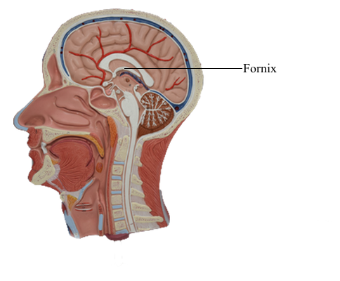
Axons of hippocampal neurons converge to form a prominent
bundle that arches around caudal, superior, and rostral aspects of
the thalamus. This bundle, the fornix, is a major efferent path of
the hippocampal formation. It is composed of a flattened caudal part, the crus; a compact superior
portion, the body; and a part that arches around the rostral part
of the thalamus and passes through the hypothalamus to terminate in the mammillary body - this is the column of the fornix.
Located along the edge of the dentate gyrus and continuing on
the lateral edge of the crus and body of the fornix is a thin fringe
of fibers called the fimbria.
The amygdaloid nuclear complex (commonly called the
amygdala) is located internal to the cortex of the uncus. It is composed of several cell groups,
including caudomedial, basolateral, and central subdivisions.
Two major efferent bundles are related to the amygdala. First,
the stria terminalis follows a looping trajectory that shadows, in
a reverse direction, the orientation of the caudate nucleus. In the temporal horn, the stria terminalis is located
just medial to the tail of the caudate nucleus. As the stria terminalis arches superiorly and rostrally, it assumes a position in
the shallow groove between the caudate nucleus and the dorsal
thalamus, where it is accompanied by the terminal vein (superior thalamostriate vein). At about the level of
the interventricular foramen, the fibers of the stria terminalis
fan out to enter and terminate in the hypothalamus, the septal
area, and the neostriatum.
The second major efferent bundle of the amygdala is the diffusely arranged ventral amygdalofugal pathway. These fibers
leave the amygdaloid complex, pass medially through the substantia innominata, and continue medially to enter hypothalamic and septal nuclei or turn caudally and distribute to the
brainstem.
Cell groups located internal to the subcallosal area collectively
form the septal nuclei. Consequently, the subcallosal
area, together with a small strip of cortex located adjacent to the lamina terminalis, the paraterminal gyrus, is commonly called the
septal area. The septal nuclei are medially adjacent to the nucleus
accumbens and continuous with sheets of neuronal cell bodies that
extend into the septum pellucidum. The latter structure extends,
in general, from the fornix to the inner surface of the corpus callosum. It forms the medial wall of the anterior horns and a small
part of the bodies of the lateral ventricles.
In general, the septal nuclei have complex interconnections with
hippocampal, amygdaloid, and other limbic structures.